
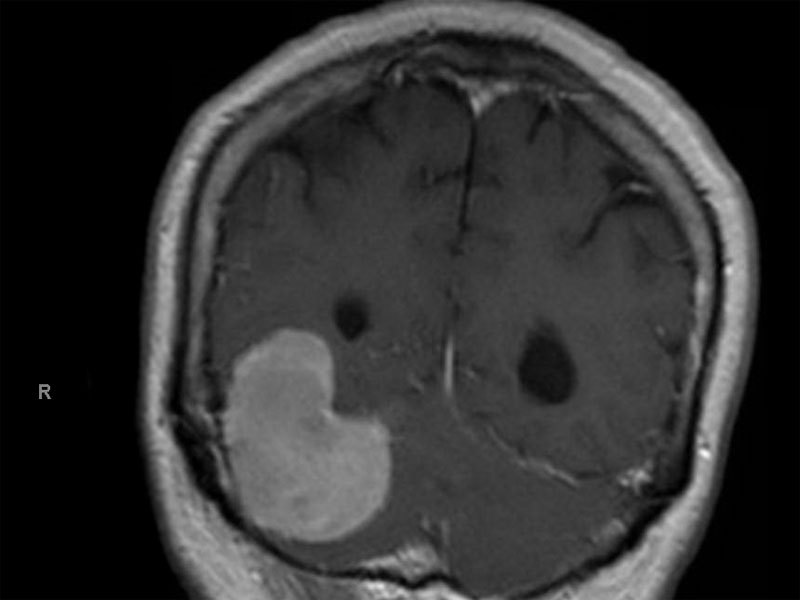
MRI shows an extra-axial lesion in the right posterior fossa with extension into the posterior right temporal lobe. There is associated mass effect with partial effacement of the fourth ventricle. These features are compatible with a meningioma.
)
A moderately cellular tumor composed of bland cells with a swirling architecture and indistinct cell borders.
)
Monomorphic cells are arranged in lobules, separated by fibrous septae.
)
Note the prominent whorling of cells. The nuclei are oval with delicate even chromatin
)
The nuclei are almost pallisading, reminisent of a schwannoma (one of the main entities on the differential). Intranuclear pseudoinclusions are present.
)
There are numerous subtypes of meningiomas, some of which demonstrate more a epithelial phenotype (e.g. microcystic, secretory, clear cell, chordoid, papillary) or more mesenchymal differentiation (e.g. fibrous, angiomatous, metaplastic)(Fletcher).
The subtypes which most closely exhibits classic "meningothelial" morphology are the meningotheliomatous (syncytial) and transitional (mixture of syncytial and fibrous subtypes) variants.
Histologically, meningiotheliomatous meningioma is composed of whorls of cells separated by a fibrous stroma. The cells have oval to round nuclei and the cell borders are indistinct (i.e. syncytial). Intranuclear pseudoinclusions (actually cytoplasmic inclusions) are common (Prayson).
This variant is classified as a WHO Grade I tumor.
• Meninges : Meningioma, Transitional Variant
• Meninges : Meningioma, Fibrous Variant
• Meninges : Meningioma, Meningotheliomatous (Syncytial) Variant
• Meninges : Meningioma, Angiomatous Variant
• Meninges : Meningioma, Chordoid Variant
• :
• Meninges : Meningioma, Microcystic Variant
• Meninges : Meningioma, Psammomatous Variant
• Meninges : Meningioma, Secretory Variant
Fletcher CDM, ed. Diagnostic Histopathology of Tumors. 3rd Ed. Philadelphia, PA: Elsevier; 2007: 1707-1710.
Kumar V, Abbas AK, Fausto N. Robbins and Cotran Pathologic Basis of Disease. 7th Ed. Philadelphia, PA: Elsevier; 2005: 1409-1410
Prayson, RA. Neuropathology: Foundations in Diagnostic Pathology. Philadelphia, PA: Elvesier; 2005: 489-494.
Prayson R, Kleinschmidt-Demasters BK, Cohen ML. Brain Tumors. Consultant Pathology Series New York, NY: Demos Publishing: 2010: 190-2.