
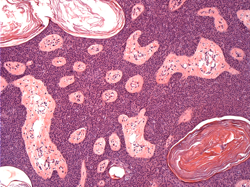
Seborrheic keratoses are characterized by epidermal proliferation which can take the form of acanthosis, papillomatosis, hyperkeratosis, horn pseudocysts (which are actually papillomatosis cut tangentially, but connect to the surface), melanin in the basal layer, basaloid appearance keratinocytes, and a sharp demarcation from the base of the epidermal proliferation to the dermis (Rapini). Epidermal hyperplasia of basaloid keratinocytes and horn pseudocysts are key features of this benign lesion seen here.
)
Seborrheic keratoses (SKs) are very common benign growths that have a "stuck-on" appearance and can appear anywhere on the skin except for palms, soles and mucosal surfaces. They arise usually after age 30 and increases in frequency with age. They carry the unfortunate name of "aging barnacles" (Busam, Rapini).
These benign lesions are not associated with sebaceous glands, however, they are so named because they commonly arise in areas with sebaceous glands (face, trunk).
Most common on the face and trunk, with a stuck on, warty, red/pink/brown appearance. More frequent as one ages -- "aging barnacle". If there are multiple eruptions of irritated SKs, it is called the sign of Leser-Trelat and may indicate a GI malignancy (Busam).
Busam KJ. Dermatopathology: Foundations in Diagnostic Pathology 1st Ed. Philadelphia, PA: Elsevier; 2010: 336.
Rapini RP. Practical Dermatopathology. Philadelphia, PA: Elsevier; 2005: 234-5.