System: CNS: Cerebellum: Metastatic: Hemangioblastoma

System: CNS: Cerebellum: Metastatic: Hemangioblastoma

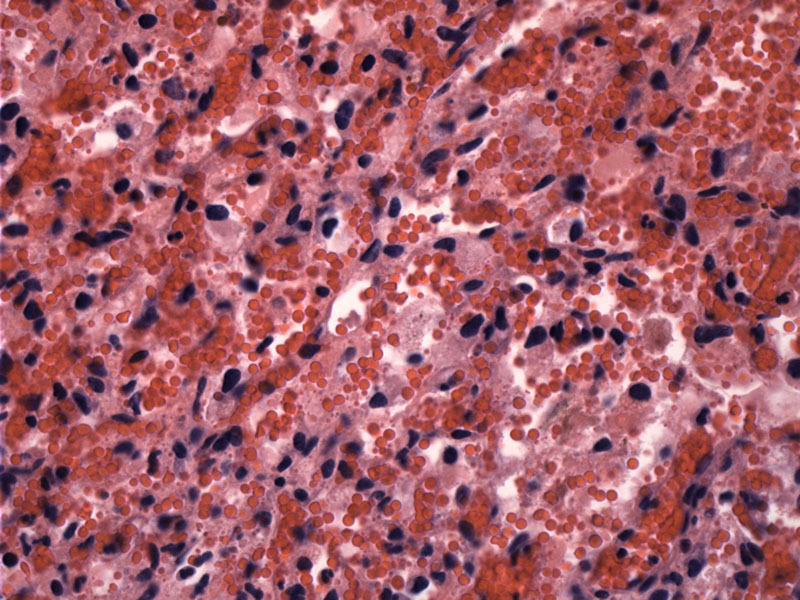
HMB is a vascular lesion composed of proliferating capillaries and stromal cells.
The tumor is characterized by a proliferation of vascular channels and stromal cells. The stromal cells can have eosinophilic or lipid-laden vacuolated cytoplasm (Bush). Nuclear pleomorphism may be present, but is not prognostically significant. This particular area consists mostly of stromal cells, which can have eosinophilic or vacuolated cytoplasm.
CD34 highlights the proliferating endothelial cells.
1 Image
2 Image
3 Image
4 Image
Hemangioblastoma, a benign vascular neoplasm, occurs almost exclusively in the central nervous system. Favored locations include the cerebellum and spinal cord. Occurrence in other CNS sites such as the supratentorial compartment, optic nerve, peripheral nerves or soft tissues of the extremities have been reported, but are extremely rare (Slavin).
There is a well known association with von Hippel-Lindau disease, which is an autosomal dominant syndrome resulting from mutation in a tumor suppressor gene located on chromosome 3p25-26. Patients with VHL have a variety of tumors including hemangioblastomas of the cerebellum (also known as Lindau tumors), angiomas of the retina, renal cell carcinoma and pheochromocytomas. Approximately 1/4 of patients with HMB have von Hippel-Lindau disease.
Grossly, the tumor is well-circumscribed, cystic with red-brown (vascular) mural nodules.
Note that rosenthal fibers may be seen in the tumor periphery and biopsies in this area may appear similar to pilocytic astrocytoma. It is important to distinguish HMB from metastatic renal cell carcinoma, which may also afflict patients with VHL (Prayson).
HMBs are twice as common in men as women (Slavin). Although these tumors can develop at any age, peak incidence occurs between the third and fourth decades. Patients with VHL develop these tumors at an earlier age (Prayson). Interestingly, these tumors also elaborate an erythropoietin-like substance that leads to secondary polycythemia; this occurs in approximately 10% of cases (Prayson, Slavin).
HMB is a WHO Grade I tumor and considered a benign lesion.
• Meninges : Meningioma, Angiomatous Variant
Bush ML, Pritchett C, Packer M et al. Hemangioblastoma of the cerebellopontine angle. Arch Otolaryngol Head Neck Surg. 2010 Jul;136(7):734-8.
Prayson, RA. Neuropathology: Foundations in Diagnostic Pathology. Philadelphia, PA: Elvesier; 2005: 496-9.
Slavin KV. Hemangioblastoma: eMedicine. Last updated on 24 March 2009. Available at: emedicine.medscape.com/article/250670-overview
)
)
)
)
)
)
)