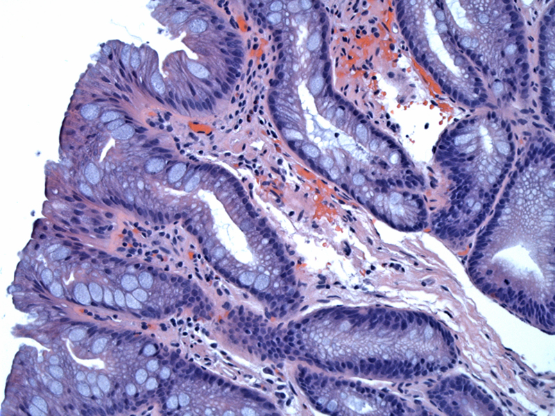

This specialized intestinal metaplastic columnar epithelium is identified by a columnar epithelium with goblet cells. This image shows goblet cells, blue columnar, clear columnar, and enterocyte-like cells. Not seen here, but sometimes scattered Paneth cells can also rarely be seen. It is now generally agreed that the goblet cell is the key cell type for the identification of Barrett's epithelium.
Barrett's epithelium is a form of metaplasia in which the normal squamous lining is replaced by a glandular lining of the specialized intestinal type. Over the past 50 years, the definition of Barrett's esophagus has gone through three major phases.
Initially the surgeon Norman Barrett described red glandular mucosa in the distal esophagus of some patients that he assumed to be a portion of the stomach pulled into the distal esophagus as a result of the scarring following ulceration.
Subsequently, three types of glandular epithelium were identified as characteristic of Barrett's esophagus: (1) specialized intestinal epithelium (with goblet cells), (2) junctional epithelium (or cardia-antral type), without goblet cells, and (3) fundic or oxyntic epithelium, also without goblet cells.
The current definition began with the recognition that specialized columnar epithelium was not a normal finding in the distal esophagus, and that only this type of epithelium was associated with an increased risk for the development of adenocarcinoma.
Barrett's esophagus may be classified as ultra long segment (at least 3 cm in length), short segment (involving < 3 cm), and ultra-short-segment (not visible endoscopically, but histologically confirmed columnar epithelium).
Barrett esophagus results from long-standing gastroesophageal reflux and occurs in up to 10% of patients with GERD. The incidence is highest in middle-aged white males. It is the most important risk factor in the development of esophageal adenocarcinoma. On endoscopy, Barrett epithelium is seen as extensions (tongue-like) of salmon-colored mucosa up from the gastroesophageal junction (Kumar).
Although the connection between Barrett's esophagus and the development of adenocarcinoma is well defined, with overall risk estimates varying from 1/52-441 patient years, or a lifetime risk for an individual patient of about 5%, the risk of carcinoma development for short-segment or ultra-short-segment Barrett's is less well defined.
• Esophagus : Gastroesophageal Reflux Disease (GERD)
• Esophagus : Indefinite for Glandular Dysplasia
• Esophagus : Glandular Dysplasia, Low Grade
• Esophagus : Glandular Dysplasia, High Grade
Kumar V, Abbas AK, Fausto N. Robbins and Cotran Pathologic Basis of Disease. 7th Ed. Philadelphia, PA: Elsevier; 2005: 804-5.