
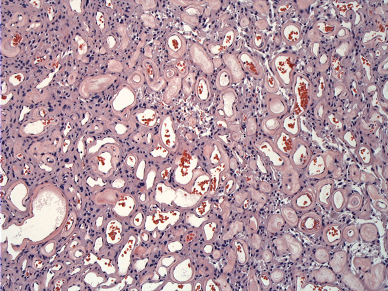
At low power, it appears the tumor is dominated by medium sized vessels.
)
The vessels are so numerous in some areas, there are little intervening meningiothelial cells, thus almost obscuring the nature of the lesion. Vessels are surrounded by a hyalinized eosinophilic coat.
)
This area has a more solid area with meningiothelial tumor cells surrounding a larger vessel.
)
The angiomatous areas here (left) transitions into a more cellular focus (right).
)
Some nuclei are slightly enlarged and appear dark. Mild pleomorphism can be present, but there are no other concerning features such as increased mitotic activity, necrosis, prominent nucleoli, etc. The differential diagnosis for this variant includes capillary hemangioblastoma, another neoplasm with vascular proliferation.
)
An example of a larger vessel with a prominent hyalinized acellular rim is seen
)
Ki67 labeling demonstrates the low mitotic activity of this variant, consistent with a WHO Grade I tumor.
)
EMA stains the meningothelial cells but not the vessels themselves.
)
Meningiomas show dual epithelial and mesenchymal differentiation, and thus stain with both epithelial and mesenchymal markers. The fibrous, angiomatous and metaplastic subtypes can be considered the "mesenchymal variants" (Fletcher).
The fibrous variant is composed of spindled cells arranged in fascicles. Whorls around vessels and psammoma bodies are common as well. The metaplastic variant exhibits focal mesenchymal differentiation such as bone, cartilage, and adipose tissue.
The angiomatous variant (demonstrated here) contains an inordinate amount of blood vessels against a background of usual meningioma features (Prayson). The larger vessels often have hyalinized walls.
The percentage of the vascular component necessary to designate the meningioma as the angiomatous variant has not been defined, but in a study of 38 angiomatous meningiomas, Hasselblatt et al. used at least 50% of the total tumor area as a criteria. Either way, this is probably not critical since this variant (unlike clear cell, chordoid, papillary or rhabdoid) does not connote a worse prognosis. Rather, it is a WHO Grade I tumor.
• Meninges : Meningioma, Chordoid Variant
• Meninges : Meningioma, Microcystic Variant
• Meninges : Meningioma, Meningotheliomatous (Syncytial) Variant
• Meninges : Meningioma, Clear Cell Variant
• Meninges : Meningioma, Fibrous Variant
• Meninges : Meningioma, Psammomatous Variant
• Meninges : Meningioma, Secretory Variant
• Meninges : Meningioma, Transitional Variant
• Cerebellum : Hemangioblastoma
Fletcher CDM, ed. Diagnostic Histopathology of Tumors. 3rd Ed. Philadelphia, PA: Elsevier; 2007: 1707-1710.
Hasselblatt M, Nolte KW, Paulus W. Angiomatous meningioma: a clinicopathologic study of 38 cases. Am J Surg Pathol. 2004 Mar;28(3):390-3.
Prayson, RA. Neuropathology: Foundations in Diagnostic Pathology. Philadelphia, PA: Elvesier; 2005: 489-494.