
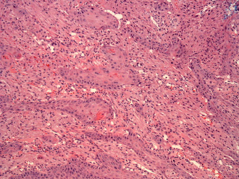
Pseudoepitheliomatous hyperplasia of epithelium obscures the underlying tumors cells, which are round lightly eosinophilic cells with granular cytoplasm and small dark nuclei with occasional nucleoli.
)
The granular cells of this lesion are seen surrounding squamous pearls and may be overlooked on first glimpse.
)
Because of their blandness, the differential can be broad and include granular variants of melanoma, leiomyomas, rhabdomyomas and other soft tissue tumors. Fortunately, granular cell tumors stain strongly for S-100 and do not stain with epithelial, endothelial, smooth muscle, melanocytic or dendritic cell markers (2).
Affects middle-aged adults with a female predilection (M:F is 2:3). Presents as slow growing painless nodules, usually on the skin or tongue, but may be found in the mucosa or submucosa of the viscera (GI tract, biliary tract, gallbladder, larynx). Approximately 5% of granular cell tumors occur in the GI tract and 1% occur in the esophagus (1,2).
Excision
Benign. Granular cell tumors are rare in general and malignant ones are even rarer. Malignant tumors would require wide resection (en bloc); radiation and chemotherapy have not proven to be effective (2).
• Ovary : Transitional Cell Carcinoma
(1)Foundations in Diagnostic Pathology
(2)eMedicine http://emedicine.medscape.com/article/282430-overview