System: Gynecological: Ovary: Neoplastic: Borderline Serous Tumor with Positive Nodes

System: Gynecological: Ovary: Neoplastic: Borderline Serous Tumor with Positive Nodes

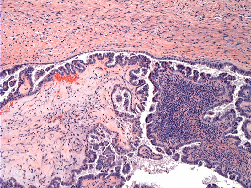
edge of the tumor, growing into a the tumor mass at the bottom. Image
tumor is papilary and lined by 1-3 cell layer thick with mild to moderate atypia Image
This pelvic node shows tumor in the subcapsular sinus region; some purple psammomatous bodies are present. There is no desmoplastic tumor response in the node, which is typical in the majority of cases. Image
Simple small papillary appearing tumor clusters are seen percolating along admixed with psammoma bodies. Image
The majority of affected nodes include the pelvic, followed by mesenteric/omental, paraaortic, and supradiaphragmatic. There is no correlation with the site of nodal involvement and overall or disease-free survival (McKenney).
Follow-up data (ranging from 8 to 230 months) on patients with nodal involvment yields overall survival and disease-free survival of 92% and 82%, respectively (McKenney). Five died of disease and 10 developed recurrent disease.
McKenney JK, Balzer BL, Longacre TA. Lymph node involvement in ovarian serous tumors of low malignant potential (borderline tumors): pathology, prognosis, and proposed classification. Am J Surg Pathol 2006. 30(5): 614-624.
)
)
)
)