
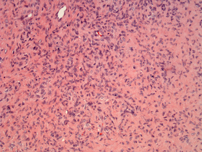
Case 1: Fibroblasts with somewhat blunt-shaped nuclei and abundant pink cytoplasm are arranged in short fascicular pattern. The fibroblasts produce the collagenous pink stroma. Although not well demonstrated in this image, slit-like vascular spaces are also characteristic of this tumor.
)
Case 2: This case is paucicellular with more extensive collagenization.
)
Stellate nuclei are embedded within a fibrocollagenous background.
)
Fibroma of the tendon sheath is a benign nodular lesion attached to a tendon sheath. It most commonly arises in the hands and feet of middle-aged men.
Microscopically, these lesions are lobulated and are composed of spindled fibroblasts against a dense fibrous background. Fibromas of tendon sheath can be differentiated from fibromatosis by their slit-like vessels and well-circumscribed gross appearance (Fletcher).
The clinical history is usually that of a slow-growing lesion. Men are twice as likely to be affected. These lesions tend to be some (less than 2 cm), well-circumscribed and lobulated. A third of patients experience some tenderness and pain. The most common sites are the tendons or tendon sheaths of fingers, hands, wrist. Sometimes, the lower extremities (knees, feet) may be affected (Wu).
Can recur if incompletely excised. No malignant potential.
Fletcher CDM, ed. Diagnostic Histopathology of Tumors. 3rd Ed. Philadelphia, PA: Elsevier; 2007: 1545.
Wu JM, Montgomery E. Classification and Pathology. Surg Clini N Am 88 (2008) 483-520.