System: Lower Respiratory : Lung: Normal: Histology

System: Lower Respiratory : Lung: Normal: Histology

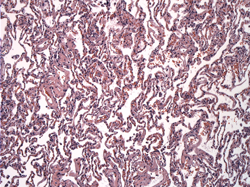
Normal alveolar parenchyma with alveolar sacs, blood vessels, and alveoli.
Higher power of normal alveolar parenchyma.
The lungs are supplied with blood from two systems. The pulmonary system carries deoxygenated blood from the right side of the heart via a large pulmonary artery to each lung. The pulmonary capallaries and surrounding alveoli are supplied by this system. The bronchial system constitutes the systemic circulation of the lower respiratory tract. It supplies blood to the lungs airway wall tissue and pleura (outer surface of the lungs). The conducting portion of the lung conditions inhaled air. Goblet cells and mucous glands produce mucus, which forms a mucous layer in most conducting tubes. The moist mucosa humidifies the air, while the mucus and ciliated epithelium filter and clean the air. The cappillary network warms air before it reaches the respiratory portion in the lungs. There are two major cells within the lung. Type 1 alveolar cells are thin simple squamous cells that line the alveoli in the lung. They are the main sites of gaseous exchange and form a blood-air barrier for respiration. Interalveolar septums are located between adjacent alveoli. The great alveolar cells are the second cell type. They are fewer in number and cuboidal in shape. They are located adjacent to Type 1 alveolar cells, within the alveoli. These cells synthesize and secrete pulmonary surfactant, a phospholipid-rich product. Pulmonary surfactant stabilizes the alveolar diameters, facilitates their expansion, and prevents their collapse. The great alveolar cells also function as stem-cells for Type 1 alveolar cells, and are believed to have some bactericidal effects which counteract dangerous pathogens. The cartilage found in the major structures of the lung are hyaline cartilage. This cartilage surrounds the intrapulmonary bronchus, along with pseudostratified columnar ciliated epithelium. The intrapulmonary bronchus consists of thin lamina propia, smooth muscle, submucosa, bronchial glands, hyaline cartilage plates, and adventitia. The branches breakdown to form smaller bronchi and bronchioles (terminal bronchiole). The terminal bronchiole do not contain cartilage plates, bronchial glands or goblet cells. These bronchiole are the smallest passageways for conducting air. They give rise to the thinner respiratory bronchioles, which are characterized by walls with numerous alveoli. These give rise to an alveolar duct which continues into the alveolar sacs. The respiratory bronchioles represent the transition zone between the conducting and respiratory portions of the respiratory system. They give rise to alveolar ducts into which open numerous alveoli. The alveolar contain macrophages and the great alveolar cells mentioned earlier. The alveolar macrophages are monocytes which clean the alveoli of invading microorganisms and inhaled particles, through a process called phagocytosis.
Eroschenko, Victor P. "diFiore's Atlas of Histology with Functional Correlations" 1oth ed, 2005. Lippincott Williams & Wilkins. Baltimore, MD.(300-306) Young, Barbara and Heath, John W. "Wheater's Functional Histology a text and colour atlas" 4th ed. 2000, Harcourt Publishers Limited. Harcourt Place, London (222)
)
)