
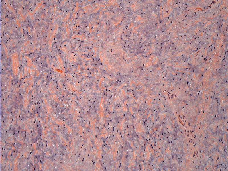
Stellate and spindled cells (seen only as dark wispy nuclei) are embedded in a loose blue myxoid stroma admixed with bands of sclerosis. There is variable cellularity, but overall, the tumor is fairly paucicellular.
)
Prominent vasculature is a constant feature, with vessels composed of small to large caliber arteries, veins, arterioles, and venules distributed among the neoplastic cells. Some large vessels showed thickened walls.
)
The large vessels (left of center and upper right) show medial hypertrophy and a branching pattern.
)
There is some hyalinization around vessel wall of this large vessel (center). Again, note the abundant myxoid stroma with interspersed fibrous areas.
)
A closer view reveals bland-appearing spindle and stellate-shaped cells with indistinct cellular borders, loosely dispersed in the myxoid matrix.
)
There are rare cells that display very minimal atypia. Mitotic activity should be scant or absent.
)
A few areas with small aggregates of plasma cells (arrows) are seen.
)
This particular case was both ER and PR positive. Idrees performed ER and PR studies on four cases of male aggressive angiomyxomas and found that one case was ER+/PR+, two cases were ER-/PR+ and one case was ER-/PR-.
)
SMA (left) and desmin (right) are both negative. One may wish to perform these stains in addition to others in order to differentiate AA from myxoid soft tissue tumors including superficial angiomyxoma, angiomyofibroblastoma (AMFB), myxoid neurofibroma and myxoid liposarcoma (Idrees).
)
According to a search conducted by Idrees (2006), there are approximately 150 cases of aggressive (deep) angiomyxomas in the vulva and 39 cases in the male inguinoscrotal region reported in the literature. The most common location is the scrotum, followed by the inguinal region.
Similar to their vulvar counterparts, testicular aggressive (deep) angiomyxomas are nonencapsulated gelatinous masses with infiltrative borders. These tumors can attain enormous sizes, and a tumor as large as 57 cm has been reported in a woman (Idrees). Characteristic features include spindle or stellate cells within a myxoid stroma with a prominent vascular component consisting of thin or thick walled vessels. The vessel walls are commonly hyalinized (Ulbright).
The initial clinical impression is inguinal hernia or testicular neoplasm, followed by spermatic cord lipoma, hydrocele, and spermatocele (Idrees).
Excision with wide margins is important in preventing recurrence. In males, the recurrence rate (9%) is much lower than that in women (30 to 40%)(Idrees, Nucci). This is attributed to anatomy -- it is much easier to completely excise a tumor in the scrotal region than in the vulva!
• Vulva : Deep (Aggressive) Angiomyxoma
Idrees MT, Hoch BL, Wang BY, Unger PD. Aggressive angiomyxoma of male genital region. Report of 4 cases with immunohistochemical evaluation including hormone receptor status. Ann Diagn Pathol. 2006 Aug;10(4):197-204.
Ulbright TM, Amin MB, Young RH. Tumors of the Testis, Adnexa, Spermatic Cord, and Scrotum: Atlas of Tumor Pathology Third Series, Fascicle 25. Washington DC; AFIP: 1999: PAGE 262-4.