
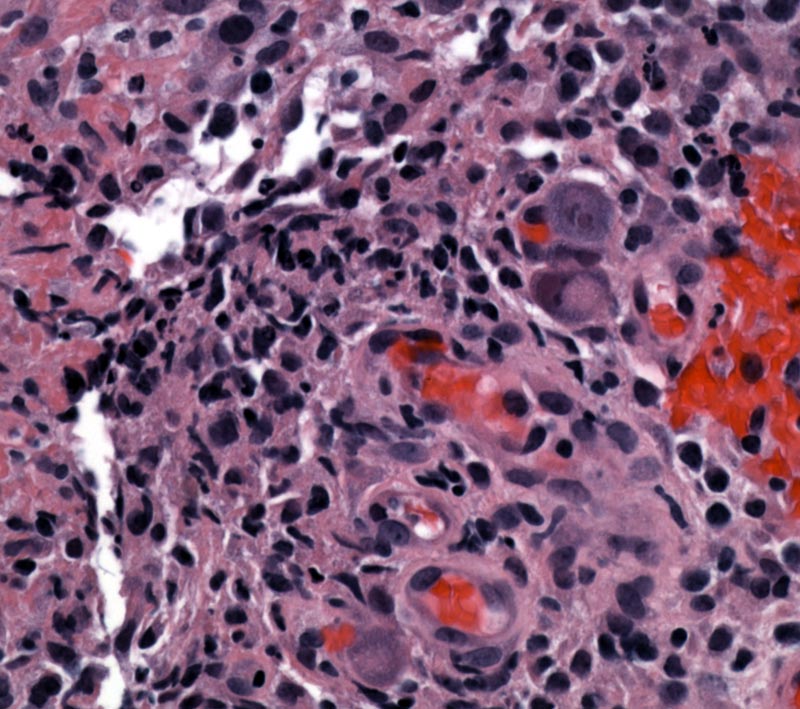
CMV colitis typically produces ulceration of the mucosa, as illustrated by the presence of a fibrinopurulent cap (left side of image). Other manifestations of exuberant infection (not shown here) can include cryptitis, crypt abscesses, crypt atrophy and numerous apoptotic enterocytes.
)
CMV cytopathic changes include cellular enlargement with virions studding the cytoplasm, as well as enlargement and smudginess to the nucleus, which is eccentrically placed. Classically, CMV inclusions are described as "owl eyes" (dark circle within a larger lighter circle), however, in many cases, you will have to use your imagination. These inclusions are found in both the nucleus and cytoplasm, and most likely to be found in the ulcer bases (versus at the edge of the ulcer like herpes inclusions). Furthermore, the inclusions are preferentially located in the endothelial and stromal cells (vs. herpes inclusions which are found in epithelial cells).
)
Most often occurs in immunocompromised patients and presents with bloody or watery diarrhea, abdominal pain, fever and weight loss. If the virus affects endothelial cells of the GI tract, it may cause ischemia (1). Patients with pre-existing chronic GI conditions such as ulcerative colitis or Crohn's disease can be superinfected with CMV, which can be fatal.
Anti-viral agents
In an immunocompetent person, infections are benign and self-limited; but in immunocompromised patients, CMV infections cause significant morbidity and mortality.
(1)Foundations in Diagnostic Pathology