
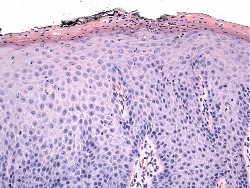
The vulvar squamous epithelium is markedly thickened and underlies a hyperkeratotic layer.
)
Neutrophils are present within the epithelium as well as within the parakeratosis.
)
PAS stain highlights a few fungal hyphae (arrow)
.', 'ihc', ''))
Vulvovaginitis is a common gynecologic condition that clinically presents with discharge, burning, vulvar irritation and itching. Common causes include bacterial vaginosis, Trichomonas and candidiasis. Vulvovaginal candidiasis is caused by the fungi Candida. The most frequently isolated species is Candida albicans, but note that there is an increasing of other specieis such as Candida glabrata, Candida tropicalis and Candida krusei. This is important to remember as certain anti-fungal agents are not as effective toward non-albicans species (Samra).
A pelvic exam reveals erythema of the vestibule and labia. Thrush patches may be found on the vulva. A thick curd-like discharge is classic . A wet mount performed on scrapings with KOH may reveal hyphae.
Topical antifungal preparations are available over the counter. Oral antifungal courses are also prescribed, with longer courses and suppression therapy for recurrent infections.
While most symptomatic patients do experience relief, recurrences do occur.
Samra-Latif OM. Vulvovaginitis: eMedicine. Last updated on April 30, 2009. Available at: emedicine.medscape.com/article/270872-overview