
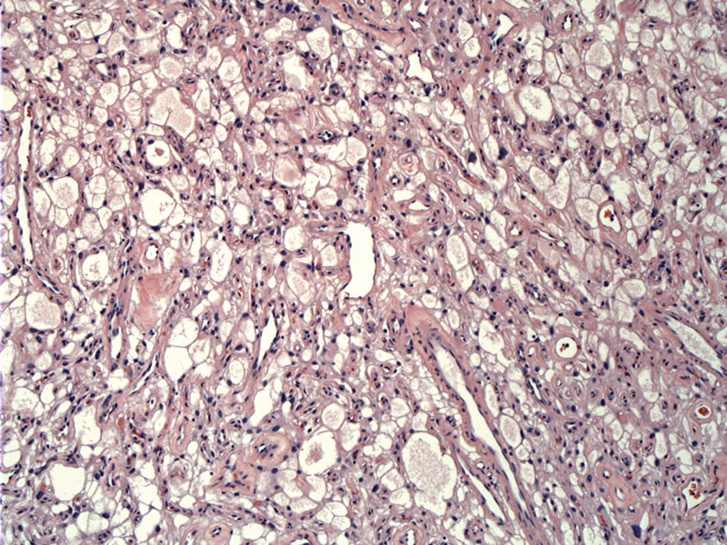
Extensive microcytic change is seen with smaller and medium sized cysts admixed.
)
The tumor shows a loose texture and microcysts with formation of extracellular spaces containing edematous fluid. The tumor cells have stellate and vacuolated cytoplasm with long cytoplasmic processes. Some nuclear pleomorphism is appreciated.
)
An angiomatous area is seen to the left blending into the microcystic architecture to the upper right.
)
Development of a cystic appearance is not only due to accumulation of extracellular fluid, but also to cytolysis due to hydropic degeneration.
)
EMA is positive as in the usual meningioma.
)
This variant is composed of areas with elongated processes and areas with a loose, mucinos background yielding an almost "swiss cheese" appearance. Loss of desmosomes accounts for the microcystic change. Classic meningioma areas are often also found. Hyalinization and nuclear pleomorphism may be encountered.
Poor enhancement on CT and MR imaging is found which may be a useful neuro-imaging characteristic indication of this variant of meningioma.
The behavior of this variant is similar to those of meningiomas in general (Nishio). The tumor is categorized as WHO Grade I.
• Meninges : Meningioma, Transitional Variant
• Meninges : Meningioma, Fibrous Variant
• Meninges : Meningioma, Chordoid Variant
• Meninges : Meningioma, Meningotheliomatous (Syncytial) Variant
• Meninges : Meningioma, Angiomatous Variant
• Meninges : Meningioma, Clear Cell Variant
• Meninges : Meningioma, Psammomatous Variant
• Meninges : Meningioma, Secretory Variant
Nishio S, et al. Microcystic meningioma: clinicopathological features of 6 cases. Neurol Res. 1994 Aug;16(4):251-6.