System: Soft Tissue: Myogenic: Neoplastic: Myofibroma

System: Soft Tissue: Myogenic: Neoplastic: Myofibroma

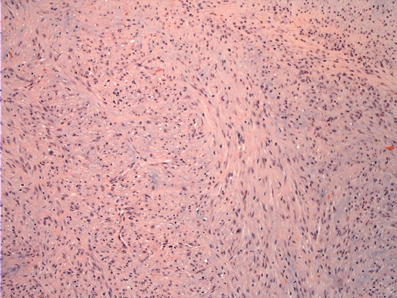
plump myoid cells in broad bundles Image
variation in cellularity with lighter staining area to the left; due to hyalinization Image
more cellular area with polygonal cells with slightly hyperchromatic nuclei Image
bundles seen, may raise the ddx of nodular fasciitis Image
irregular shaped vessel is typical; note some lymphocytes in background Image
hemangiopericytoma like vessels are typical Image
Myofibromas and myofibromatosis are a group of lesions originally thought to be congenital, but have now been described in all ages.
They can be single lesions (solitary myofibroma) or multiple (myofibromatosis). Regardless of their centricity and the age of the patient, these lesions tend to favor the head and neck region. In a study of 79 myofibromas/myofibromatosis conducted by Foss and colleagues in 2000, the mean age was 26 years. The most frequently affected site was the mandle, followed by tongue, lips and buccal area. 1/3 of the tumors affected the bones of the jaws, about half of these tumors were intramedullary (intraosseous), and all of these patients were younger than 18.
Foss RD, et al. Myofibromas and myofibromatosis of the oral region: A clinicopathologic analysis of 79 cases. Oral Surg Oral Med Oral Pathol Oral Radiol Endod 2000;89-57-65.
Mentzel T, et al. Myopericytoma of Skin and Soft Tissues: Clinicopathologic and Immunohistochemical Study of 54 Cases. Am J Surg Pathol 2006;30:104-113.
)
)
)
)
)
)