System: Soft Tissue: Myxoid: Neoplastic: Myxofibrosarcoma, High Grade

System: Soft Tissue: Myxoid: Neoplastic: Myxofibrosarcoma, High Grade

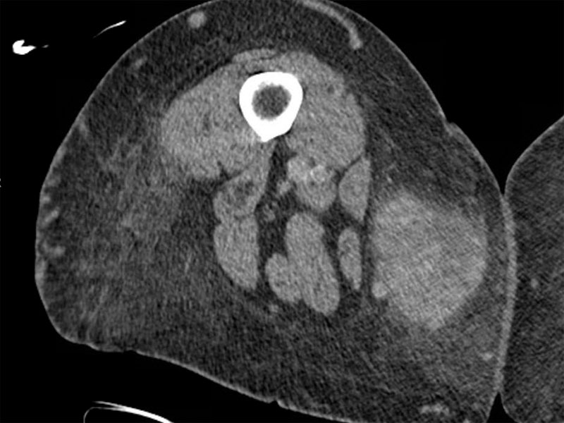
This CT scan reveals a 7cm mass in the distal medial thigh measuring. The lesion is located in the subcutaenous fat and does not appear to involve the musculature. The resection was sent to surgical pathology, with a diagnosis of myxofibrosarcoma (high grade).
gross thigh mass with hemorrhage and necrosis Image
cellular and atypia Image
pleomorphism; i have addiional images to upload for this tumor Image
Previously referred to as "mxyoid malignant fibrous histiocytoma", the WHO 2002 edition currently recognizes mxyofibrosarcoma (MFS) as a distinct entity. This tumor tends to arise in the extremities of the elderly.
Histologically, MFS exhibits the following features: (1) myxoid background (2) arcuate capillaries (3) nuclear atypia and (4) variable presence of pseudolipoblasts.
The lesions are grade from I to III depending on cellularity. Grade I lesions are hypocellular with minimal nuclear atypia whereas grade III lesions are hypercellular with more pronounced nuclear atypia. Metastatic potential increases with grade (Willems). Unlike other myxoid neoplasms on the differential (e.g. myxoid liposarcoma and low grade fibromyxoid sarcoma), there is no specific chromosomal aberration seen in myxofibrosarcoma. Complex cytogenetic abnormalities are seen in all grades of the lesion (Willems).
The lesion generally presents as a slow-growing mass in the extremity of an older individual. In a study of 158 patients (median age 64) with MFS, 61% were located in the lower extremities, 24% in the upper extremities, 15% in the trunk and the remaining 1% in the head and neck.
Sanfilippo et al (2010) found that patients with MFS have a better prognosis compared to other sarcoma patients. In their study of 158 patients with MFS, the overall five year survival was 77%. The 5 year local recurrence rate was 18% and distant metastases occurred in 15%. The higher local recurrence rate was attributed to the multinodular growth particular of MFS, making it very difficult to achieve clear margins during resection.
Important prognostic factors included size, histologic grade and resection margins. Those higher grade lesions prefer to metastasize to lungs, bone and lymph nodes.
• Lipomatous : Liposarcoma, Dedifferentiated Type
• Lipomatous : Liposarcoma, Myxoid Type
Sanfilippo et al. Myxofibrosarcoma: Prognostic Factors and Survival in a Series of Patients Treated at a Single Institution. Ann Surg Oncol (2011) 18:720-5.
Willems SM, et al. Local recurrence of myxofibrosarcoma is associated with increase in tumor grade and cytogenetic aberrations, suggesting a multistep tumor progression model. Modern Pathology (2006)19, 407-416.
)
)
)
)