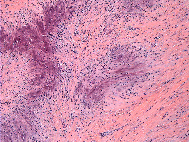

Irregular islands of calcification are surrounded by plump sometimes radially oriented fibroblast type cells.
Here is a calcified area with surrounding plump fibroblastic cells. You can see why these fibroblasts are sometimes described as chondroblastic.
These epithelioid fibroblasts have round vesicular nuclei. Pleomorphism and mitotic activity are not features of CAF.
Here is more spindled area, consisting of a proliferation of fibroblasts.
Infiltration into adjacent fat and other surrounding tissue is common and the reason for frequent recurrence following local excision.
Foci of cartilagenous metaplasia can be seen here. Longstanding lesions can be overtly cartilaginous.
Multinucleated giant cells resembling osteoclasts can be present in some cases.
Calcifying aponeurotic fibroma (CAF) presents as a slow growing painless mass in the hands (especially palms) or feet. Most arise in children and adolescents (age 8-14) with a male predilection. In rare instances, these lesions may arise more proximally i.e. thigh or trunk.
Radiographs reveal a mass usually with calcific stippling. Grossly, these masses are infiltrative and merge with the surrounding soft tissues and tendon. On cut section, gritty areas represent calcified areas.
Histologically, there is a proliferation of spindled fibroblasts. The unique feature of CAF is that areas of calcification and chondroid matrix are surrounding by radially oriented and/or palisading fibroblasts that exhibit a more epithelioid, plump areas. Occasionally, osteoclastic giants cells and ossification can be present. Pleomorphism and necrosis are not seen.
Typically occurs in young children and adolescents with a male predilection. Presents as a soft tissue mass in the hand or wrist (Rosai, Fletcher). At excision, these masses are infiltrative and often attached to tendons.
Conservative excision is recommended. As CAF is often adherent to dense fibrous connective tissue (eg, tendon, fascia, or periosteum), it may not be possible to excise the tumor entirely, especially since most cases arise in the hand.
Recurrence is not infrequent due to its infiltrative margins. Exceptionally rare cases of malignant transformation into sarcoma has been documented (Folpe).
→Areas of calcifications surrounding by radially oriented, palisading plump almost chondroblastic fibroblasts are the key histologic features.
→Mostly occurs on the palms or wrists of young children or adolescents.
Fletcher CDM, ed. Diagnostic Histopathology of Tumors. 3rd Ed. Philadelphia, PA: Elsevier; 2007: 1549.
Folpe AL, Inwards CY. Bone and Soft Tissue Pathology: Foundations in Diagnostic Pathology Philadelphia, PA: Elsevier; 2010: 51-2.
Rosai, J. Rosai and Ackerman's Surgical Pathology. 9th Ed. Philadelphia, PA: Elsevier; 2004: 2243.
