
Extensive coagulative necrosis (bottom) is common in undifferentiated carcinoma.
)
Undifferentiated single and some giant cells contain a moderate amount of lightly eosinophilic cytoplasm. Bizarre mitotic figures are scattered throughout.
)
The pleomorphic cells have markedly hyperchromatic nuclei, and there is surrounding dissecting hemorrhage. Again note the brisk mitotic activity.
)
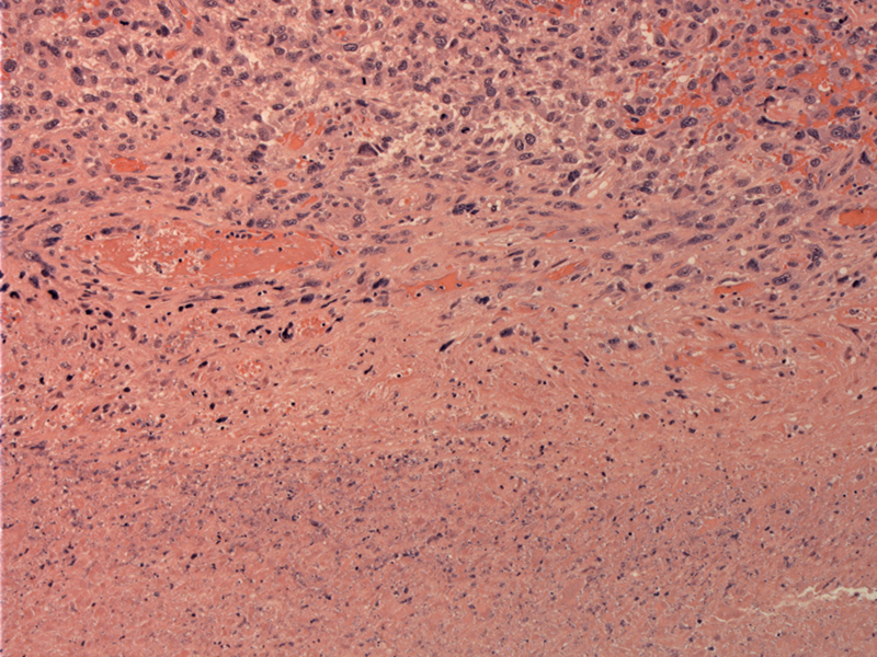
A solid sheet of tumor cells (bottom) with geographic necrosis (top) is typical.
)
Undifferentiated (anaplastic) carcinomas of the thyroid usually affect the elderly. The clinical presentation is usually that of a fixed firm mass that has invaded local structures and metastasized to regional lymph nodes and/or lung. The prognosis is dismal and most survive only 6 months. It is thought the the tumor arises from dedifferentiation of a pre-existing well-differentiated papillary carcinoma, however, there are a subset of tumors that arise de novo.
Histologically, the tumor is composed of spindle cells, pleomorphic giant cells and epithelioid cells. One cell type may predominate. In this case, the tumor was largely composed of pleomorphic giant cells.
• Thyroid : Anaplastic Carcinoma, with Osteoclast-like Giant Cells
• Thyroid : Anaplastic Carcinoma, Spindle Cell Type
• Thyroid : Anaplastic Carcinoma, with Osteoclast-like Giant Cells
• Thyroid : Anaplastic Carcinoma, Squamoid Type
DeLellis RA, Lloyd RV, Heitz PU, Eng C. Tumors of Endocrine Organs: WHO Classification of Tumours. Lyon; IARC Press; 2004: 77-80.
Fletcher CDM, ed. Diagnostic Histopathology of Tumors. 3rd Ed. Philadelphia, PA: Elsevier; 2007: .
Thompson LDR. Endocrine Pathology: Foundations in Diagnostic Pathology. Philadelphia, PA: Elsevier; 2006: 109-113.