
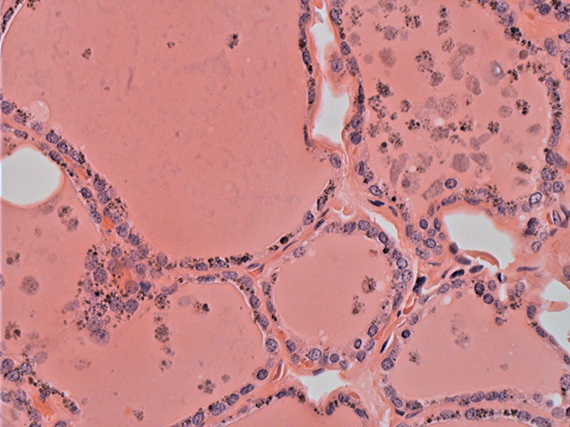
Follicular epithelial cells contain some coarsely granular dark brown pigment.
)
In addition, pigmented macrophages are found within the colloid.
)
Long term use of minocycline, a long-acting antibiotic of the tetracycline group, can result in dark brown to black pigment accumulating in the thyroid. The composition of the pigment is not clear, nor is the mechanism of altered deposition. The pigment accumulates within the lysosomes of the follicular epithelial cells (Bell, Saul).
Approximately 30 cases of black thyroid had been reported in the English literature thus far (Tsokos). Most cases go unrecognized and are discovered when the thyroid is removed for other reasons. Almost all published cases show normal function of the thyroid. The thyroid appears diffusely black when removed.
→Upon seeing a black thyroid grossly, think of minocycline use.
→The pigment accumulates in the follicular cells and colloid.
Bell CD, Kovacs K, Horvath E, Rotondo F. Histologic, immunohistochemical, and ultrastructural findings in a case of minocycline-associated "black thyroid". Endocr Pathol. 2001 Winter;12(4):443-51.
Saul SH, Dekker A, Lee RE, Breitfeld V. The black thyroid. Its relation to minocycline use in man. Arch Pathol Lab Med. 1983 Apr;107(4):173-7.
Tsokos M, Schröder S. Black thyroid: report of an autopsy case. Int J Legal Med. 2006 May;120(3):157-9. Epub 2005 Sep 6.