System: Skin: Epidermis: Neoplastic: Basal Cell Carcinoma, Superficial Type

System: Skin: Epidermis: Neoplastic: Basal Cell Carcinoma, Superficial Type

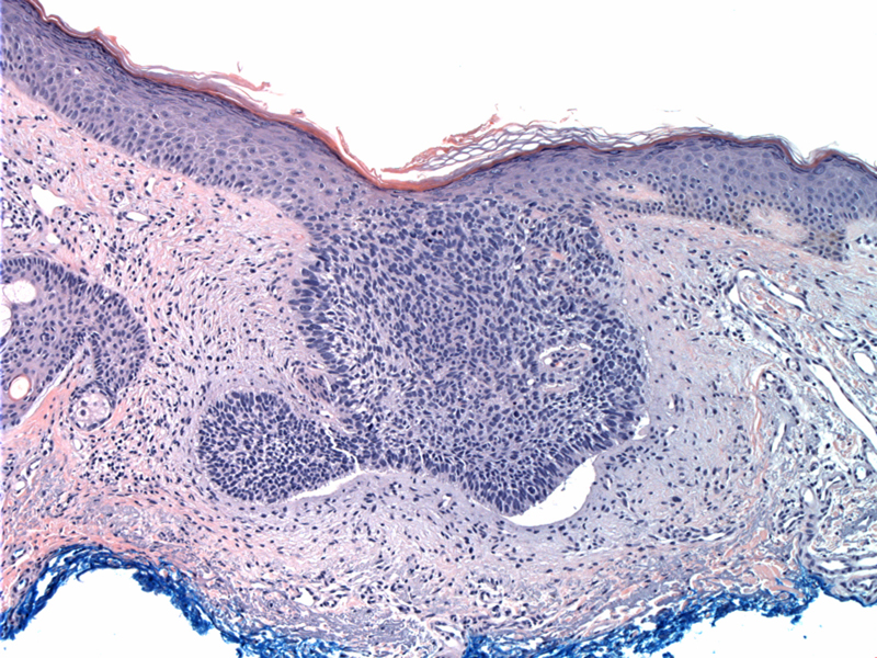
Case 1: A proliferation of atypical basaloid cells appears to bud off from the epithelium but still remains attached to it. Note the slit-like retraction of the palisaded basal cells from the subjacent myxoid stroma.
Superficial BCCs are characterized by epithelial aggregates of basaloid cells that are still attached to the epidermis. These aggregates are multifocal and areas of normal epidermis (skip areas) are in between the proliferative nests (Busam).
In a study of 175 BCCs, the superficial subtype occurred more often in younger patients and on the trunk. In contrast, nodular BCCs preferentially affected the head and neck region of older patients. Morpheaform BCCs also preferentially occurred in the head and neck region and the prevalence did not differ between the younger and older age groups (Betti).
superficial BCC subtype occurs more often at a younger age, particularly in females. This variant often manifests itself as a reddish plaque with variegate depigmentation and a spreading peripheral margin and/or an atrophic or scar-like peripheral border that raises clinical concern for Bowen's disease
• Epidermis : Basal Cell Carcinoma
Betti R, et al. Anatomic location and histopathologic subtype of basal cell carcinomas in adults younger than 40 or 90 and older: any difference? Dermatol Surg. 2009 Feb;35(2):201-6.
Busam KJ. Dermatopathology: Foundations in Diagnostic Pathology 1st Ed. Philadelphia, PA: Elsevier; 2010: 389-397.
)