System: Skin: Epidermis: Neoplastic: Basal Cell Carcinoma, Infiltrating Type

System: Skin: Epidermis: Neoplastic: Basal Cell Carcinoma, Infiltrating Type

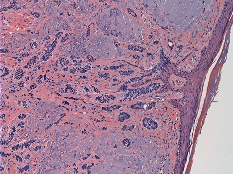
Case 1: This image clearly demonstrates its origin from the epidermis. Note the sun damaged skin (solar elastosis).
There is deep involvement down into the underlying fat. Columns of basaloid cells one to two cells thick are enmeshed in a densely collagenized stroma.
Cords of jagged irregular basaloid cells within a desmoplastic stroma is the typical appearance.
Case 2: Another case that was also deeply infiltrative into the deep dermis and subcutis. Nests show sharp angulation of their peripheral contours; there is frequent mitotic activity and individual cell necrosis.
Here is a more solid area with irregular outlines and marked desmoplasia; peripheral pallisading is not present.
Morpheaform BCC and infiltrative BCCs are characterized by irregularly-shaped and angulated aggregates of basaloid cells, arranged in strands and small nests. Single cells percolating through a fibroplastic stroma is also common. There is still slit-like retraction, but less frequent than that seen in the superficial and nodular types (Crowson).
These aggressive subtypes to penetrate deeply into the dermis and subcutis; perineural invasion is a risk. Note that morpheaform BCC is synonymous with sclerosing BCC and are associated with prominent stromal fibrosis. However, infiltrative BCC may not necessarily be morpheaform or sclerosing (Busam).
The infiltrative and morpheaform/sclerosing types present clinically as white or yellow depressed fibrotic scars that rarely ulcerate or bleed, and are most often on sun exposed skin. These variants must be distinguished from other desmoplastic adnexal tumors such as desmoplastic trichoepithelioma (presence of papillary mesenchymal bodies and horn cysts) and microcystic adnexal carcinoma (presence of sweat ducts and horn cysts).
Regarded as more aggressive due to its tendency to be deeper; higher rate of local recurrence.
• Epidermis : Basal Cell Carcinoma
• Adnexal : Desmoplastic Trichoepithelioma
• Adnexal : Microcystic Adnexal Carcinoma
Busam KJ. Dermatopathology: Foundations in Diagnostic Pathology 1st Ed. Philadelphia, PA: Elsevier; 2010: 389-397.
Crowson AN. Basal cell carcinoma: biology, morphology and clinical implications. Modern Pathology (2006) 19, S127-S147.
)
)
)
)
)