
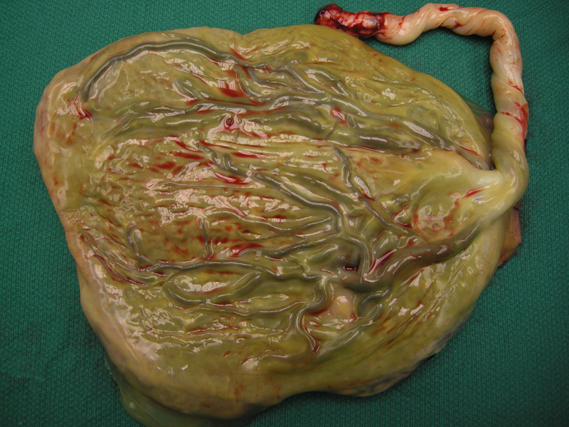
Heavy meconium staining involves the membranes in this placenta, resulting in an apparent green-tinged appearance.
)
Pigmented macrophages (round cells with a slight brown tinge) contain meconium in this section of fetal membrane.
)
Brown tinged cytoplasm corresponds to placental uptake of meconium.
)
Approximately 13% of live births show meconium-stained amniotic fluid. Green-colored fetal membranes are frequently the result of meconium staining but may also be imparted by changing blood pigments from an earlier bleeding event or by myeloperoxidase in leukocytes in infections. Thick green slime that easily clears off the membranes is consistent with meconium (Walsh). Meconium is thought to be passed in utero in times of fetal stress.
Typically pediatricians will be present at the time of delivery in the event of meconium passage to assist in agressive suctioning of the oropharynx and possible the trachea.
In addition to signifying antepartum fetal stresses, meconium aspiration is a significant neonatal complication that may predispose newborns to neonatal repiratory distress syndrome.
Walsh MC, Fanaroff JM. Meconium stained fluid: approach to the mother and the baby. Clin Perinatol. 2007 Dec;34(4):653-65.