
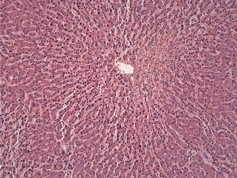
In this CMV infected liver, necrosis ia centered primarily on the central vein.
)
The areas of necrosis are becoming confluent and forming bridges.
)
Viral inclusions cannot be identified, but discrete islands of cell death can be seen.
)
Here is a microabscess, with neutrophils surrounding an infected cell.
)
Cytomegalovirus (CMV) hepatitis is most commonly seen in immunocompromised patients, and CMV is the most important infectious organism in solid organ transplant patients (Iacobuzio). The infection is usually due to reactivation of a latent infection, but primary infections can occur as well. The new infection may be acquired from blood transfusions.
CMV infection is usually self-limited with mild symptoms, however, in immunocomprised patients, CMV infection can be deadly. Microscopically, characteristic owl-eye viral inclusions can be seen in hepatocytes, endothelial cells and bile duct cells. These inclusions are not always present, but one should see microabscesses which surround the infected cells, consisting of neutrophils with varying amounts of mononuclear inflammatory cells (Iacobuzio, Cheng). IHC studies for CMV will help highlight infected cells.
Microabscesses and owl-eye inclusions are helpful diagnostic features for CMV hepatitis.
• Lymph Nodes : CMV Lymphadenitis
• :
• Liver : Herpes Simplex Hepatitis
Cheng L, Bostwick DG, eds. Essentials of Anatomic Pathology. 2nd Ed. Totowa, NJ: Humana Press; 2006: 1377.
Iacobuzio-Donahue CA, Montgomery EA. Gastrointestinal and Liver Pathology: Foundations in Diagnostic Pathology. Philadelphia, PA: Elsevier; 2005: 544-5.