System: Mediastinum: Thymus: Neoplastic: Thymic Carcinoma, Squamous Type

System: Mediastinum: Thymus: Neoplastic: Thymic Carcinoma, Squamous Type

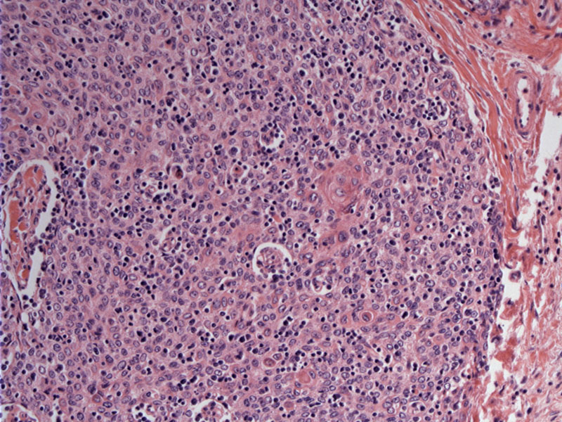
This is an example of a well-differentiated squamous cell carcinoma subtype. Epithelial cells with abundant light pink cytoplasm form a well-demarcated nodule. Note the squamous pearl just right of center.
The mildly pleomorphic cells have vesicular nuclei and prominent nucleoli. There is a sparse infiltrate of lymphoid cells.
Look closely -- you can even see intercellular bridges.
The squamous cell subtype of thymic carcinoma is the most common histologic variant. It is further subdivided into (1) well-differentiated or keratinizing, (2) moderately differentiated and (3) poorly differentiated (nonkeratinizing). Note that some poorly differentiated forms are often accompanied by a lymphoplasmacytic infiltrate and some authors prefer to label them as the "lymphoepithelial-like carcinoma". About 50% of these lesions are associated with EBV infection (Suster, Fletcher). We will discuss lymphoepithelial-like subtype in a separate case.
The neoplastic infiltrate is virtually identical to squamous cell carcinoma of the lung, thus, it is important to distinguish between the two because thymic carcinoma (squamous type) usually carries a much more favorable prognosis (Fletcher, Suster).
Histologically, there is a vague lobular pattern where lobules of neoplastic cells are separated by fibrous and desmoplastic stroma. In well-differentiated tumors, the neoplastic epithelial cells will exhibit keratinization and intercellular bridges (Fletcher).
Fletcher CDM, ed. Diagnostic Histopathology of Tumors. 3rd Ed. Philadelphia, PA: Elsevier; 2007: 1331-2
Moran CA, Suster S. Thymic carcinoma: current concepts and histologic features. Hematol Oncol Clin North Am. 2008 Jun;22(3):393-407.

)
)
)