
Fragments of squamous epithelium from the esophagus show hyperplasia of the basal zone, even accounting for tangential sectioning. Morphologic features of GERD include basal zone hyperplasia (normal is a 3 cell layer),elongation of dermal papillae (extending greater than 2/3 of the thickness of the epithelium), spongiosis of the epithelilum layer, increase in intraepithelial lymphocytes, eosinophils and neutrophils.
)
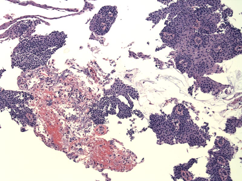
A soft feature of GERD is the presence of red cell congestion. Image
)
The epithelium shows intercellular spongiosis along with a marked increase in the number of intervening mature lymphocytes, also known as squiggle cells.
)
This better oriented section shows basal zone hyperplasia, elongation of the rete, and intraepithelial eosinophils. While an occasional eosinophil may be acceptable in the non-pediatric population, when present in these numbers and accompanied by the other features of GERD, they are helpful in making the diagnosis. Again note the red cell congestion in the papillary areas.
)
The most important cause of esophagitis is reflux of gastric contents into the lower esophagus, usually as a result of decreased LES (lower esophageal sphincter) tone. There are many other predisposing factors such as a sliding hiatal hernia, scleroderma and pregnancy.
Mucosal injury results from the chronic assault of gastric juices, and in severe cases, bile from the duodenum. As a side note, in esophageal pH monitoring, detection of a pH less than 4 (for a specified amount of time within a 24 hour period) suggests reflux from the stomach, but detection of a pH greater than 7 suggests alkaline reflux of bile from the duodenum.
Presents as heartburn, dysphagia and regurgitation of sour-tasting gastric material.
Medications (H2-recepter antagonists and proton-pump inhibitors) or in difficult cases, surgery (fundoplication and/or reduction of hiatal hernia).
Complications include progression to intestinal metaplasia (Barrett's), bleeding, ulceration and fibrosis leading to the development of a stricture.
• Esophagus : Barrett Esophagus (Intestinal Metaplasia)
1 Kumar V, Abbas AK, Fausto N. Robbins and Cotran Pathologic Basis of Disease. 7th Ed. Philadelphia, PA: Elsevier; 2005: 804
2 Rosai, J. Rosai and Ackerman's Surgical Pathology. 9th Ed. Philadelphia, PA: Elsevier; 2004: 619-20.