
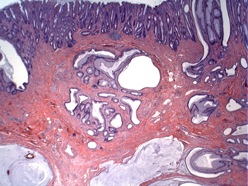
The surface epithelium is shown in the uppermost part of the image, while the submucsal contains displaced glands arranged in a somewhat lobular pattern with jagged borders.
)
The submucosa contains calcification and cystic spaces often devoid of epithelium. Pools of mucin raise the question of invasive mucinous adenocarcinoma, so one must be careful to assess this finding in the context of the entire histological impression.
)
The collections of glands show a pushing front into underlying stroma with intervening fascicles of smooth muscle.
)
Mucinous cystic spaces are lined by epithelium which may be attenuated or denuded in some areas. In contrast to mucinous adenocarcinoma, the epithelium in colitis cystica profunda does not exhibit atypia.
)
Colitis cystica profunda is a rare condition involving the colonic submucosa characterized by mucus-filled cysts with a benign epithelial lining. There is a localized form (also known as hamartomatous inverted polyp) which is usually located in the rectum and related to solitary rectal ulcer syndrome. The diffuse form arises from inflammation and ulceration of the mucosa and is seen in ulcerative colitis, Crohn disease and irradiation change (Rosai).
It is important to recognize this rare lesion because the finding of submucosal mucous-containing glands may lead to the misdiagnosis of invasive mucinous adenocarcinoma of the rectum. However, the epithelium lining the glands in colitis cystica profunda does not exhibit atypia.
Patients of a wide age range (children to elderly) are potentially affected. Blood in the stool, mucoid stools, or diarrhea are typical presenting signs.
High fiber diet may alleviate symptoms, but some require surgical resection due to obstruction, or to exclude adenocarcinoma.
• Colon : Inflammatory Cloacogenic Polyp
Iacobuzio-Donahue CA, Montgomery EA. Gastrointestinal and Liver Pathology: Foundations in Diagnostic Pathology. Philadelphia, PA: Elsevier; 2005: 366.
Rosai, J. Rosai and Ackerman's Surgical Pathology. 9th Ed. Philadelphia, PA: Elsevier; 2004: 798.