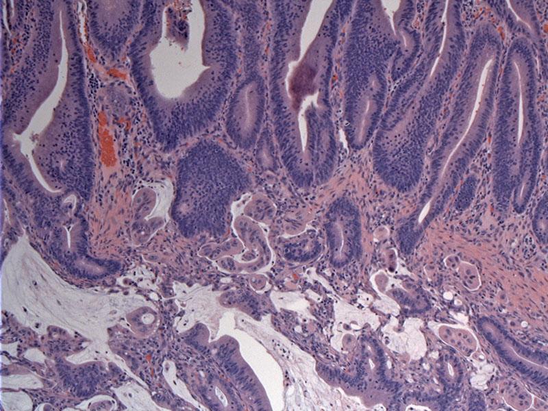

The invasive component (lower image) directly arises from a surface villous adenoma (upper image).
The duodenal surface is involved by villous adenoma with elongated projections composed fibrovascular cores and overlying adenomatous tall columnar epithelium.
While not typical, this particular intestinal type adenocarcinoma is comprised largely of extracellular mucin pools. Malignant epithelium is seen in association with the mucin as the tumor dissects into the muscularis propria.
This particular tumor is unusual because most that arise do not contain this much extracellular mucin.
The tumor focally invades the pancreatic duct, as shown here.
The ampulla is covered by two types of epithelium which can give rise to adenocarcinoma. Therefore, either intestinal type adenocarcinoma (the more common type) or the pancreaticobiliary type may arise in this location. The intestinal type looks like the usual adenocarcinoma arising in the GI tract. The pancreaticobiliary type resembles ductal adenocarcinoma from the pancreas or bile ducts.
Some conditions such as familial adenomatosis polyposis (FAP), celiac disease, Crohn's, and Lynch syndrome are associated with an increased incidence of small intestinal carcinoma.
Usually affects those in their 60s-70s but are seen in younger patients with a hereditary cancer syndrome. Symptoms may include intussescption, bleeding, or obstruction.
• Small Intestines : Adenocarcinoma with Mucinous Features