System: Breast: Ductal: Malignant: Ductal Carcinoma In situ


endocrine variant - this stained strongly for synapto and chromogranin
This image demonstrates high grade DCIS with comedo necrosis. Note the neoplastic cells in the periphery exhibit pleomorphism. High grade DCIS tends to have a solid pattern with central comedo necrosis. Note, however, that comedo necrosis can also occur in other patterns such as micropapillary and cribriform.
This micropapillary and cribriform low grade pattern Image
tufting and micropapillary low grade dcis Image
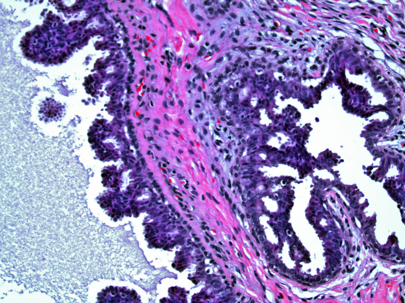
another nice example of micropapillary pattern Image
LG DCIS involving a complex papillary lesion Image
Ductal carcinoma in situ (DCIS) is a neoplastic proliferation of epithelial cells in the terminal ductal lobular unit (TDLU). DCIS is considered a precursor to invasive ductal carcinoma, although not every case of DCIS will progress to invasive cancer. The relative risk of invasive carcinoma if DCIS is found is approximately 10.0 to 11.0 (please check).
DCIS is typically classified by nuclear grade (high, intermediate, low) with a comment on the architectural pattern (cribriform, micropapillary, solid). Lesions often demonstrate a mixture of patterns. Note that "comedo" necrosis, signifying the presence of central necrosis within a proliferation of malignant cells is merely a descriptor, and not a subtype. Thus, the term "comedo DCIS" does not provide information regarding nuclear grade nor an architectural pattern.
DCIS is identified mammographically as microcalcifications, or less commonly, DCIS may present as Paget's disease or a breast mass.
• Lobular : Lobular Carcinoma in Situ, Pleomorphic Variant
)
)
)
)
)
)
)