
The laryngocele consists of a cystic space resulting from abnormal dilatation of the laryngeal saccule, which rests between the ventricular folds, the base of the epiglottis and the inner surface of the thyroid cartilage. Image
)
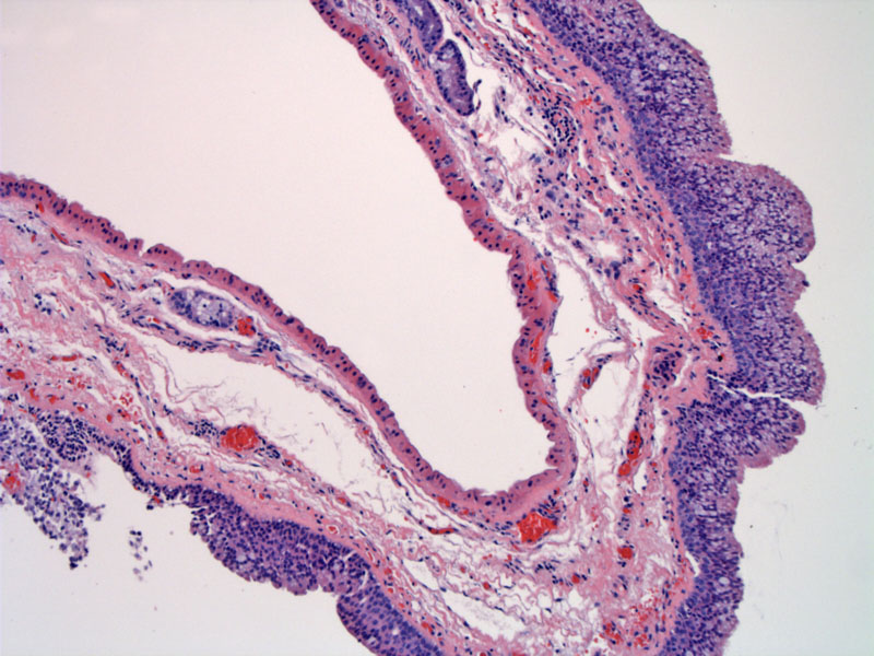
It may be lined by a variety of epithelial types -- in this example there is bland oncocytic epithelium. Respiratory type epithelium is also common, as is squamous metaplasia.
)
A laryngocele represents a dilation of the saccule containing air, and it maintains communication with the larygneal lumen. Most are unilateral but up to 1/4 may be bilateral. There are three types: internal (dilated confined to the intrinsic larynx); external (dilated sacs extend between the false cord and thyroid cartilage and laterally through thyrohyoid ligament; and mixed. One important complication is infection.
Most commonly encountered in the 5-8th decade. Symptoms are rare, but hoarseness, coughing, dysnea may occur. There are some case reports on patients presenting with airway obstruction. May arise in those with a tendency to excessive increase intralaryngeal pressure, such as musicians, or co-exist with laryngeal squamous cell carcinoma.
Asymptomatic cases can be left alone. Large or symptomatic ones may necessitate excision.