
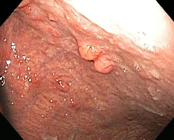
Endoscopy shows multiple small polypoid lesions. This pattern would be consistent with either type I or type II.
)
The large majority of gastric carcinoids are multiple and asymptomatic, arising in the corpus and fundus, in the setting of hypergastrinemia.
)
Inflamed surface mucosa is undermined by a proliferation of uniform round cells in orderly cords.
)
Ribboning is one pattern for carcinoid as seen here.
)
This carcinoid arose in a background of chronic atrophic gastritis with intestinal metaplasia (note goblet cells to the right at 3:00).
)
CD56 shows variably intense positivity.
)
Chromogranin is strongly positive.
)
Types of Gastric Carcinoid are summarized in this chart.
)
Carcinoid tumors (a.k.a. well-differentiated neuroendocrine tumor) are derived from resident endocrine cells in tissues. They can be found in virtually all organs, but the most common sites are the lungs and GI tract.
Gastric carcinoids originate from gastric enterochromaffin-like (ECL) mucosal cells and account for 2.4% of all carcinoids. The WHO classification has divided gastric carcinoids into three major subtypes (see chart, last image).
Type I (75%): Often small, multiple, polypoid, multicentric arising in the setting of hypergastrinemia due to chronic atrophic (autoimmune) gastritis. These patients produce auto-antibodies to parietal cells, causing their destruction. Parietal cells elaborate intrinsic factor, which aid in the absorption of vitamine B12, thus Type I patients develop pernicious anemia. The lack of parietal cells also leads to loss of negative feedback to gastric-producing cells, thus, gastrin is increased and induces the formation of carcinoids (Iacobuzio). Metastases occur in 2-5% of cases and proliferative activity (Ki-67) is <2%. Thus, most type I carincoids have benign behavior.
Type II (15%): Often small and multiple lesions arise in patient with Zollinger-Ellison syndrome in association with multiple endocrine neoplasia type I. Patients with ZES have gastrinomas that elaborate gastrin. The gastrin leads to peptic ulceration, often in unusual locations such as the jejunum. Gastrinomas and other endocrine tumors (i.e. type II carcinoid) are part of MEN I. Metastases occurs in about 10% of cases and proliferative activity (Ki-67) is <2%. Thus, most type II are considered a low-grade malignancy.
Type III (20%): These are sporadic and solitary polypoid masses, often ulcerated and larger than type I or II carcinoids. The serum gastrin level is normal. Lymph node and distant metastases occurs in more than 50% of cases and proliferative activity (Ki-67) >2%. This is considered an aggressive tumor with a mortality rate of 25%.
A type IV has been proposed in which hypergastrinemia is secondary to a primary defect of gastric acid production by parietal cells, leading to hypergastrinemia and multiple small carcinoid tumors. In summary, type I, II and IV are associated with hypergastrinemia, are usually small multiple lesions that rarely metastasize. They are located in the fundus of the stomach. Type III is NOT associated with hypergastrinemia, occur as a solitary lesion in the prepyloric region; metastasis and aggressive behavior is the norm (Fletcher).
Microscopially, uniform round cells form nests, trabeculae or glandlike structures. The tumor cells have a "salt and pepper" chromatin patter. They will be reactive for neuroendocrine markers such as synaptophysin, chromogranin A or NSE (Iacobuzio).
May present with abdominal pain, gastrointestinal bleeding, anemia and dyspepsia. Only 10% patients present with carcinoid syndrome. Note that carcinoid syndrome only occurs if the serotonin escapes degradation by the liver (e.g. liver metastases or bronchogenic in origin).
Achieving successful accuracy and the correct characterization requires extensive sampling from both the antrum (two samples) and body-fundus (four samples).
Treatment depends upon the type and size of lesions. For type I lesions, a conservative approach based on endoscopic resection is often sufficient when the size (< 1 cm) and the number (< 3-5) of the tumors renders this feasible. Management of type III lesions is comparable to that for gastric adenocarcinomas, which includes partial or total gastrectomy with extended lymph node resection.
→Type I is associated with chronic atrophic autoimmune gastritis; Type II is associated with Zollinger-Ellison syndrome; Type III are sporadic lesions and are aggressive lesions.
• Stomach : Large Cell Neuroendocrine Carcinoma
Fletcher CDM, ed. Diagnostic Histopathology of Tumors. 3rd Ed. Philadelphia, PA: Elsevier; 2007: 353-5.
Iacobuzio-Donahue CA, Montgomery EA. Gastrointestinal and Liver Pathology: Foundations in Diagnostic Pathology. Philadelphia, PA: Elsevier; 2005: 136-9.