
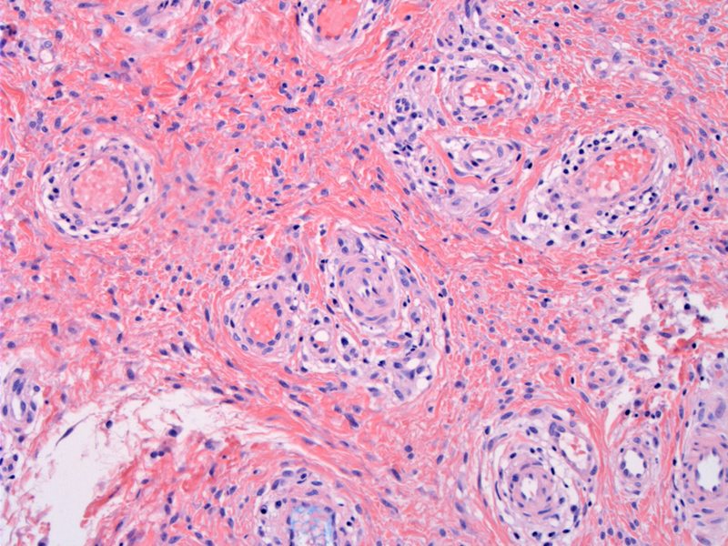
Thick-walled vessels are set in a fibrous stroma consisting of short interlacing fascicles of spindle cells. Wispy bundles of collagen are also dispersed throughout.
)
Hemangiopericytoma-like (delicate staghorn-shaped) vessels are present just under the skin surface - note the more characteristic area on the right consisting of thick-walled vessels set in a fibrous stroma.
)
Hyalinized vessels are highly characteristic. Distinction from aggressive angiomyxoma relies upon the circumscription of cellular angiofibroma and its higher cellularity, a more fibrous and less myxoid stroma, and a greater tendency for hyalinization of vessel walls.
)
Cellular angiofibromas arise in the vulvovaginal of reproductive-age and middle-age women. These tumors are also common in the inguinoscrotal regions of men and may occur in extragenital sites.1,2
Grossly, these lesions form a well-circumscribed, painless subcutaneous mass. Microscopically, as with most vulvar mesenchymal lesions, there is a stromal component and a vascular component. The stromal component consists of short intersecting fascicles of bland spindle cells. The vascular component consists of small to medium caliber vessels with thick, often hyalinzed walls. Scattered small wispy bundles of collagen are also highly characteristic.2
In terms of IHC, 60% of cases are positive for CD34, 20% are positive for SMA and 8% are positive for desmin. ER and PR may be positive in 50% of cases.1
Affects middle-age women (average age of 53.5 years). Presents as a well-circumscribed, painless subcutaneous mass that is usually less than 3cm.
Excision with adequate margins is sufficient -- these neoplasms are benign and generally do not recur.
• Vulva : Angiomyofibroblastoma
• Vulva : Deep (Aggressive) Angiomyxoma
• Vulva : Superficial Angiomyxoma
1 Fletcher CDM, ed. Diagnostic Histopathology of Tumors. 3rd Ed. Philadelphia, PA: Elsevier; 2007: 740
2 Nucci MR, Oliva Esther. Gynecologic Pathology: Foundations in Diagnostic Pathology. Philadelphia, PA: Elsevier: 2009: 36-8.