
Endoscopic image of villous adenoma
IMAGE DESCRIPTIONS
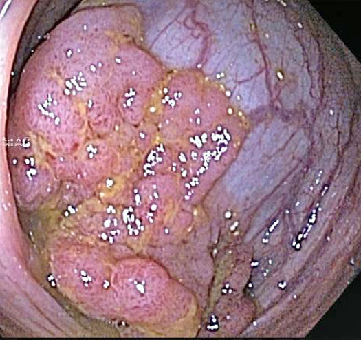
Endoscopy shows a large lesion with heaped up margins in the cecum
)
The corresponding resection from the cecum shows an exophytic lesion with heaped up edges, but proved to be a villous adenoma.
)
Adenomatous polyps of the GI tract are by definition at least low grade dysplasia. Adenomas can be either tubular, tubulovillous, or villous. At least 75% of the lesion should appear villous to qualify as as a villous adenoma.
)
Delicate finger-like structures with some underlying crypt complexity are seen here in this villouw adenoma.
)
The fibrovascular cores are lined by epithelial cells wtih pseudostratified nuclei.
)
RELATED DIAGNOSES
• Colon : Tubulovillous Adenoma
Last updated: 2010-11-16
For questions, comments or feedback on this case: editor@surgpath4u.com