
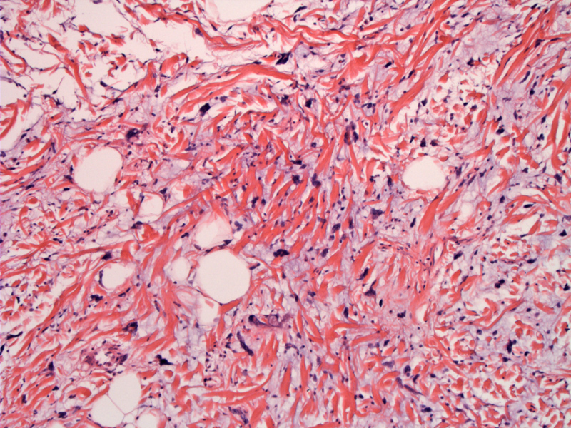
The lesion is comprised of pleomorphic and multinucleated giant cells in a myxoid stroma, along with an admixture of mature adipocytes, fibroblasts. Thick, ropy, coarse, mature, birefringent collagen bundles are distributed as well.
)
Multinucleated giant cells are either of the floret-like or nonfloret type. The floret-like multinucleated giant cells contain a moderate amounts of eosinophilic cytoplasm and peripherally arranged hyperchromatic nuclei that often overlap-- this arrangement resembles the petals of a flower. And while floret cells are a key feature in the diagnosis, they are not pathognomonic and may be seen in other lipomatous neoplasms.
)
The admixture of cells stains differently by immunohistochemical means. Tumor cells show positivity for CD34 (floret cells and fibroblasts), coagulation factor XIIIa positive (histiocytes), and weak S-100 protein positive (floret and pleomorphic cells),
)
Spindle cell and pleomorphic lipoma share common karyotypic abnormalities (rearrangements of 13q and 16q) and some overlapping histological features as well. Tumors are superficial, and therefore any such tumors arising in the deeper tissues should be regarded with suspicion.
Pleomorphic lipoma characteristically occurs in the subcutis of the neck and shoulder in men aged 50–70 years. The mass is usually is present for many years prior to diagnosis.
Treatment is complete conservative surgical excision with clear margins. Simple enucleation is insufficient due to the potential (albeit infrequent) for recurrences.
• Lipomatous : Spindle Cell Lipoma
Yencha MW, Hodge JJ. Pleomorphic lipoma: case report and literature review. Dermatol Surg. 2000 Apr;26(4):375-80.