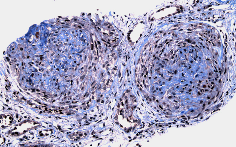

Two glomeruli evaluated on Trichrome are severely affected by cellular crescents. Note that the crescents may become fibrocellular and eventually, completely fibrotic.
There is a nearly concentric cellular crescent filling Bowman's space and beginning to compress the glomerulus on this PAS stain. The surrounding parenchyma shows scattered mature lymphocytes and some fibrosis.
Silver stain helps to delineate the crescent. The percentage of glomeruli afftected by crescents often exceeds 80%, and this percentage usually correlates to renal function as well as outcome after treatment.
Necrosis of tubules is profound. There is simplified epithelium with dilated tubules and eosinophilic material in center, which consists of necrotic tubular epithelial cells and fibrin. The interstitium contains lymphocytes with edema.
Anti-GBM disease is the result of antibodies direct against antigens in the glomerular basement membrane. This results in the characteristic linear pattern of IgG and C3 staining on immunofluorescence; in contrast, a granular staining pattern is seen in circulating immune complex deposition.
The anti-GBM antibodies can also cross-react with basement membranes in other locations such as the lung. When antibodies react with lung alveolar and glomerular basement membranes, it results in Goodpasture syndrome. The targeted antigen is the non-collagenous domain (NC1) of the alpha3-chain of collagen type IV, which is an important protein in the maintenance of the GBM architecture (Kumar).
Anti-GBM antibody-induced nephritis manifests as rapidly progressive (crescentic) glomerulonephritis. In RPGN, rapid and progressive loss of renal function occurs with oliguria and renal failure and death (if untreated). RPGN has numerous etiologies (not just anti-GBM disease), and is broadly categorized based on immune mechanism: (1) Type I RPGN is caused by anti-GBM antibodes; (2) Type II RPGN is caused by immune complexes e.g. SLE, Henonch-Schonlein purpura (IgA nephropathy); (3) Type III RPGN (pauci-immune) e.g. ANCA associated, Wegener granulomatosis.
Microscopically, there is crescent formation and glomerular fibrinoid necrosis. The crescents, composed of epithelial cells and inflammatory cells, may initially be cellular and eventually become fibrotic. Rupture of the glomerular basement membrane and Bowman's capsule may be seen, especially if highlighted by silver stain. There is usually accompanying acute interstitial inflammation and acute tubular injury (Zhou).
Many present with a reno-pulmonary syndrome -- the combination of renal and pulmonary insufficiency. However, other types of presentations include those with only renal involvement. Microhematuria afftects almost all patients, while many have macrohematuria and rapidly progressive renal insufficiency. Symptoms for those with lung involvement are hemoptysis, exertional dyspnoea, cough and fatigue.
Of note, up to 1/3 of patients with anti-GBM disease have antineutrophil cytoplasmic antibodies (ANCA), which usually associated with pauci-immune crescentic glomerulonephritis such as Wegener granulomatosis or microscopic polyangiitis. The presence of ANCA in patients with anti-GBM disease usually indicates that there is small vessel vasculitis beyond the kidney and lung (Rosai).
Plasmapheresis to remove the anti-GBM antibody and immunosuppressive therapy.
Sometimes progression is explosive leading to anuria within days, while a minority experience a protracted a course where the renal function is preserved for several months.
Kumar V, Abbas AK, Fausto N. Robbins and Cotran Pathologic Basis of Disease. 7th Ed. Philadelphia, PA: Elsevier; 2005: 968, 976-7.
Rosai, J. Rosai and Ackerman's Surgical Pathology. 9th Ed. Philadelphia, PA: Elsevier; 2004: 1197-1198.
Zhou M, Magi-Galluzzi, C. Genitourinary Pathology: Foundations in Diagnostic Pathology. Philadelphia, PA: Elvesier; 2006: 384-6.