System: Head and Neck: Sinonasal: Benign: Nasal Mesenchymal Hamartoma

System: Head and Neck: Sinonasal: Benign: Nasal Mesenchymal Hamartoma

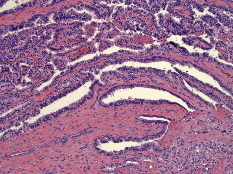
congenital lesion consisting of a proliferation of columnar type epithelium in a pseudoacinar and papillary pattern Image
admixed with fat Image
embedded irregularly in a spindled stroma Image
stroma is intensely positive for smooth muscle actin Image
focal area of glial differentation Image
confirmed by strong GFAP staining Image
papillary areas lined by bland columnar cells arranged around a fibrovascular core, are remniscent of choroid plexus Image
lower power Image
columnar cell papillary/acinar areas merge imperceptible into stroma as round cells with abundant light pink cytoplasm arranged in cords Image
glial tissue in the upper portion of image, merges with the glandular areas; fascicular bundles noted towards the left likely smooth muscle Image
cystic dilatation Image
shows surface epithelium with underlying process Image
papillary area merges with a focus resembling excessive basement membrane as seen in some salivary neoplasms Image
neural? not sure Image
)
)
)
)
)
)
)
)
)
)
)
)
)
)