System: Lower Respiratory : Lung: Neoplastic: Large Cell Carcinoma

System: Lower Respiratory : Lung: Neoplastic: Large Cell Carcinoma

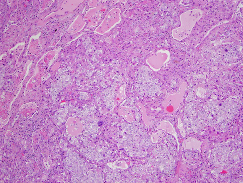
This LCC has vesicular nuclei with prominent nucleoli and abundant cytoplasm.
large dark cells, not sure about diagnosis -- no prominent nucleoli Image
ttf1 is positive in up to half Image
another area of same tumor Image
Large cell carcinoma (LCC) are comprised of large epithelial cells that do not show glandular or squamous cell differentiation. LCC used to be called "undifferentiated large cell carcinoma", however it has now been demonstrated that these tumors have immunohistochemical and ultrastructural profiles of epithelial cells, and thus, the term undifferentiated is no longer employed.
Approximately 10-20% of all lung cancers are LCC, and they tend to arise peripherally with pleural invasion (Cheng, Zander), although the basaloid variant tends to be centrally located. These tumors tend to be larger than 4 cm with necrosis. Microscopically, neoplastic cells with large nuclei and prominent nucleoli form sheets or nests. Several variants fall under the category of LCC: large cell neuroendocrine, basaloid, lymphoepithelioma-like and clear cell (Zander). In contrast to pleomorphic carcinoma, giant cells and spindle cells are not present in LCC.
LCC is strongly associated with smoking. Imaging typically shows a large peripherally located mass.
• Lung : Large Cell Neuroendocrine Carcinoma
Cheng L, Bostwick DG, eds. Essentials of Anatomic Pathology. 2nd Ed. Totowa, NJ: Humana Press; 2006: 948.
Fletcher CDM, ed. Diagnostic Histopathology of Tumors. 3rd Ed. Philadelphia, PA: Elsevier; 2007: 186.
Zander DS, Farver CF. Pulmonary Pathology: Foundations in Diagnostic Pathology. Philadelphia, PA: Elvesier; 2008: 555-7.
)
)
)
)