
First trimester villi are large and bulbous -- mostly consisting of mesenchymal villi and immature intermediate villi. The stroma is cellular and edematous.
)
At 20 weeks, this is a second trimester placenta with immature intermediate villi transitioning to stem villi. The transition is gradual and includes formation of vessels with a media and adventitial layer as well as a concomitant increase in stromal collagen.
)
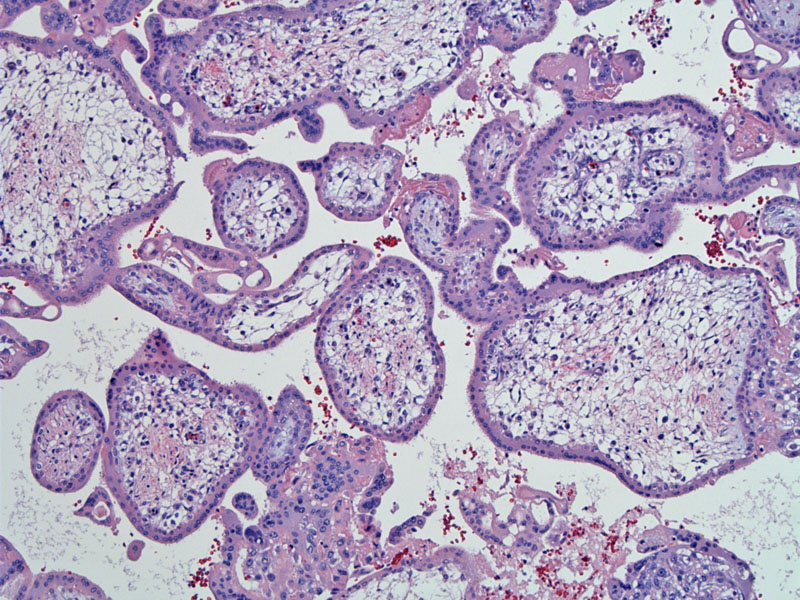
Another area of this 20 week 2nd trimester placenta demonstrates a fluid-filled cellular stroma forming reticular channels. Note the absence of vasculature.
)
Third trimester villi are much smaller and crowded as it consists largely of mature intermediate villi and terminal villi. Note the dispersed presence of syncytial knots.
)
The development of villi during gestation, if discussed in excruciating detail, can be quite confusing. The key point about villi maturation is that early in gestation, the villi are large, and edematous with a bulbous contour -- they are covered by a thick trophoblastic cell layer. As the placenta matures, there is increased branching of the villi and the villi become smaller in caliber with numerous vessels in the stroma to facilitate nutrient exchange.
In chronic placental ischemia (ie. pre-eclampsia), placenta of earlier gestational ages will exhibit hypermaturity, meaning that the villi exhibit 'mature' features such as terminal branching with syncytial knots as the placenta tries to obtain more nutrients in the setting of uteroplacental insufficiency. Now, if you are an Ob/Gyn resident you can stop here. It is enough to know that early placentas (placentae for wordsmiths) have big bulbous villi and mature placenta have small villi with numerous fetal vessels located in the periphery.
For more details on villous maturation, read on:
There are 5 main subtypes of villi, which are categorized according to their appearance during gestation, caliber/size, presence of fetal vasculature and hierarchy within the villous tree. The five types are: mesenchymal villi, immature intermediate villi, mature intermediate villi, stem villi and terminal villi. In the first and second trimesters, mesenchymal villi give rise to immature intermediate villi which later develop into stem villi. In the third trimester, mesenchymal villi give rise to mature intermediate villi, which develop into terminal villi.1
Thus, mesenchymal villi are the 'source' of all other types of villi. At term, the approximate constitution is: 40-50% terminal villi, 25% mature intermediate villi, 20=25% stem villi, 5-10% immature intermediate villi and less than 1% mesenchymal villi.1
All villi are covered by a trophoblastic shell consisting of an outer syncytiotrophoblastic layer and an inner cytotrophoblastic layer. This cover wraps around the villous mesenchyme from which the fetal vessels emerge. As secondary and tertiary villi develop in the 2nd and 3rd trimesters, several events occur: (1) the villi become smaller in caliber; (2) the outer layer of syncytiotrophoblasts becomes increasingly attenuated and form aggregates called syncytial knots; (3) the mesenchyme, edematous and cellular in early development, becomes less cellular and provides a simple support matrix for fetal vessels. Also the number of Hofbauer cells (villous macrophages) decreases in mature villi.2
Briefly, microscopic features of the five subtypes are as follows:
Mesenchymal villi: thick trophoblastic shell with prominent Langhan cells. Langhan cells are not well described in the literature, but they are not to be confused with Langerhan cells, which are dendritic cells in the skin containing Birbeck granules. Located between the syncytium and trophoblastic layer, Langhan cells are supposedly regenerative cytotrophoblasts that produce syncytiotrophoblasts. The stromal core of mesenchymal villi consists of a loose collection of collagen, fibroblasts and numerous Hofbauer histocytes. Fetal vasculature is absent.
Immature intermediate villi: Derived from mesenchymal villi, they have a bulbous contour with a thick trophoblastic cover. The stromal core is edematous and reticular, containing Hofbauer cells.
Stem villi: Derived from immature intermediate villi, they provide the support for the villous tree. Also having a thick trophoblastic shell, the stroma contains condensed fascicles of collagen. Occasional fibroblasts and macrophages are seen. In larger stem villi, there is a central muscular artery and vein. In the periphery of the villi, small vessels can be seen which feed the stem villi (similar to vasa vasorum of larger vessels in other organs).
Mature intermediate villi: Derived from mesenchymal villi in the third trimester, they are long and slender (when cut longitudinally). The stroma contains loose bundles of haphazardly arranged connective tissue fibers with numerous delicate vascular structures.
Terminal villi: Derived from mature intermediate villi, they are the terminal ends of the villous tree. There is a thin trophoblastic cover -- the stroma contains over 50% peripherally located fetal vessels, surrounded by loose connective tissue.
1 Baergen RN. Manual of Benirschke and Kaufmann's Pathology of the Human Placenta. New York, NY: Springer; 2005: 96-105.
2 Mills, S. Histology for Pathologists. 3rd Ed. Philadelphia, PA: Lippincott, Williams & Wilkins; 2007: 1113-4.