System: Cardiovascular: Vascular: Reactive: Atherosclerotic Plaque

System: Cardiovascular: Vascular: Reactive: Atherosclerotic Plaque

plaque removed from left posterior descending coronary artery shows a tubular configuration of fibrointimal hyperplasia (right) Image
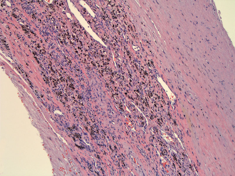
hemosiderin indicates prior hemorrhage; capillaries indicative of neovascularization formed within the plaque in an attempt to recanalize the lumen to blood flow Image
)
)