
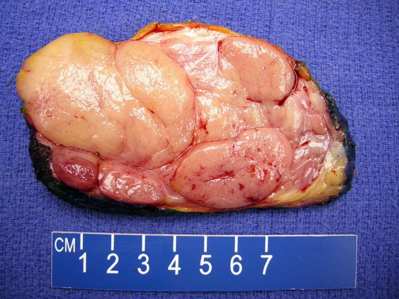
Juvenile fibroadenoma presents as a large bulging uniform tumor with a lobulated well-circumscribed nature.
)
The histologic pattern of the juvenile fibroadenomas is very similar to that of conventional fibroadenomas. Juvenile fibroadenomas generally exhibit the pericanicular growth pattern (tubular structures in a background of fibrous stroma). Cleft-like spaces of the intracanalicular pattern is not common.
)
The epithelial proliferation is usually florid and reminiscent of gynecomastia (see our case for gynecomastia). The epithelial component may also mimic atypical ductal hyperplasia. A good clue to distinguish between the two entities is that the luminal projections of juvenile fibroadenoma are slender micropapillary formations, whereas the luminal projections of atypical ductal hyperplasia are more bulbous.
)
Fibroadenomas are usually 1-3 cm in size, but in some instances (especially in younger women), they may grow rapidly and exceed 20 cm in size and thus are termed "juvenile" or in some instances "giant" or "cellular" fibroadenomas (O'Malley, Humphrey). Microscopically, these juvenile fibroadenomas have more cellular stroma and may mimic phyllodes tumor (Humphrey). However, cleft-like spaces typical of phyllodes tumor is rarely seen. Rather, the pericanalicular pattern predominates in juvenile fibroadnoma and this is a helpful discriminating feature.
This variant of fibroadenoma occurs in adolescents and characteristically exhibit rapid growth, reaching sizes of up to 20 cm in diameter. The overlying nipple and skin may become distorted. Grossly, they are very well-circumscribed.
Excision is curative.
Excellent. These tumors are completely benign and do not recur after surgery.
→Juvenile fibroadenoma, a variant of fibroadenomas, arise in adolescents and grow rapidly, exceeding 20 cm in some cases.
→Should be distinguished from atypical ductal hyperplasia and benign phyllodes tumor.
Fletcher CDM, ed. Diagnostic Histopathology of Tumors. 3rd Ed. Philadelphia, PA: Elsevier; 2007:907.
O'Malley FP, Pinder SE. Breast Pathology: Foundations in Diagnostic Pathology. Philadelphia, PA: Elvesier; 2006:116.