
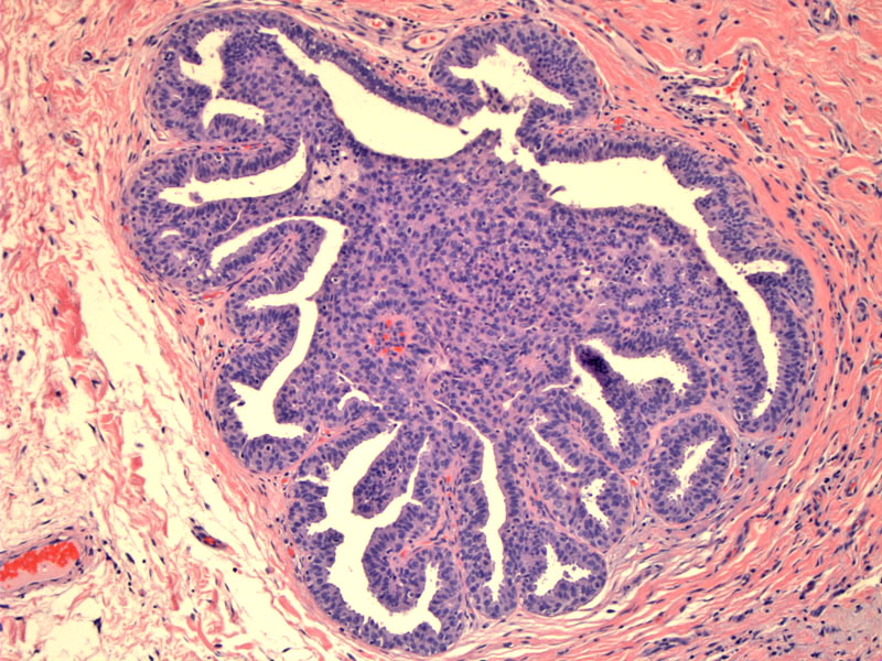
This intraductal papilloma exhibits proliferative changes of the usual type.
)
A closer look reveals streaming epithelial cells and slit-like lumina. There are no features of clonality and monotony that would be concerning for DCIS.
)
The epithelial cells lining the fibrovascular cores of a intraductal papilloma can exhibit proliferative changes. As a result of increased cellularity, the arborizing papillae with individual fibrovascular cores may be obscured. There may be solid areas of cells that form slitlike lumina.
As with UDH and DCIS, it is important to distinguish between usual hyperplasia from atypical ductal hyperplasia involving a papilloma. Cellular streaming, lack of cytologic atypia, overlapping (but bland) nuclei, inconspicuous cell borders are all clues supporting a non-neoplastic/clonal process (Ueng, Mulligan).
A solitary papilloma without atypia carries a 2 fold risk of developing breast cancer, whereas multiple papillomas (papillomatosis) without atypia carries a 3 fold risk. The presence of atypia will increase the risk to 5-7 fold (Mulligan).
• Papillary : Intraductal Papilloma
Mulligan AM, O'Malley FP. Papillary Lesions of the Breast. Adv Anat Pathol 2007;14:108-119.
Ueng SH, et al. Papillary Neoplasms of the Breast: A Review. Arch Pathol Lab Med. 2009;133:893-907.