
This particular mucinous carcinoma demonstrated largely expansile-type invasion, but there were areas of infiltrative-type invasion. Microinvasion can be seen near bottom while the more common expansile pattern can be seen in the upper right corner.
)
Another area of this tumor demonstrated closely packed glands with atypia. This is pretty typical for an expansile pattern with back to back glands with little intervening stroma.
)
Small glands appear to bud off but these are NOT invasive glands. However, note severe atypia within the glands. This area does not qualify for this diagnosis.
)
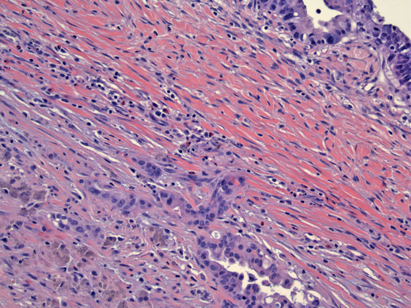
In this largely expansile-type mucinous carcinoma, there were areas suspicious for infiltrative invasion. Note the clusters of malignant cells deep into the stroma.
)
Mucinous cystadenocarcinomas are currently broadly divided into two types based on their pattern of invasion. The expansile type consists of back to back glands, with very little intervening stroma. Interobserver variability definitely exists for this diagnosis, for while some pathologists may call a mucinous carcinoma "expansile-type invasion", others may assert the same entity to be an "intraglandular carcinoma" or even "non-invasive". However, these differences are not as egregious as one would think, for intraglandular, borderline and expansile (also known as confluent) type carcinomas are usually indolent, stage I tumors that do not spread beyond the ovary (Lee).
On the other hand, the infiltrate-type pattern of invasion is characteristic of a very aggressive tumor. Most women who die from mucinous carcinomas of the ovary exhibit this pattern. Microscopically, one sees single or clusters of malignant cells percolating or dissecting through the stroma.
Note that both infiltrate-type and expansile-type carcinoma must be beyond which is considered microinvasive carcinoma. Microinvasive carcinoma is defined as one or more foci of infiltrative invasion 10 square mm or less. For the expansile pattern, the pattern of growth must also exceed 10 square mm and be at least 3 mm in each of the two linear dimensions (Lee).
Microinvasive mucinous carcinomas or those with an expansile growth pattern usually present at stage I and have a favorable prognosis with rare instances of metastasis. In contrast, mucinous carcinomas with an infiltrative growth pattern are more aggressive and seen in the majority of high-stage tumors (Hart).
• Ovary : Mucinous Cystadenocarcinoma (Expansile Type)
• Ovary : Mucinous Cystadenocarcinoma (Expansile Type)
• Ovary : Mucinous Borderline Tumor, Intestinal-type
• Ovary : Mucinous Cystadenoma
Hart WR. Mucinous tumors of the ovary: a review. Int J Gynecol Pathol. 2005 Jan;24(1):4-25.
Lee KR, Scully RE. Mucinous tumors of the ovary: a clinicopathologic study of 196 borderline tumors (of intestinal type) and carcinomas, including an evaluation of 11 cases with 'pseudomyxoma peritonei'. Am J Surg Pathol. 2000 Nov;24(11):1447-64.