
Follicles appear relatively tightly packed and are composed of cells with eosinophilic granular cytoplasm. The nuclei are round with a centrally placed distinct nucleoli (Fletcher). Occasionally, the colloid may be calcified and simulate psamomma bodies.
)
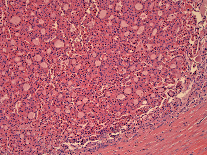
A fibrous capsule separates the adenoma from normal thyroid tissue (far right). Similar to conventional follicular neoplasms, the differentiation of adenoma from carcinoma depends on the presence of capsular or vascular invasion.
)
Variable sized follicles are lined by cells with abundant granular pink cytoplasm. The nuclei have prominent centrally placed nucleoli.
)
Hurthle (oncocytic, Askanazy) cell adenomas/carcinomas are considered variants of follicular neoplasms. Often, one sees areas of oncocytic change within a conventional follicular neoplasm. To be defined as a Hurthle cell adenoma, the tumor should be composed predominantly of oncocytic cells (Thompson, Fletcher).
Grossly, as with all oncocytic neoplasms, they have a brown hue as a result of abundant mitochondria. There is a tendency for these neoplasms to undergo infarction with hemorrhage and cyst formation after FNA (Thompson, Fletcher).
Hurthle cell adenomas are benign entities. There is some evidence that Hurthle cell carcinomas occur in older patients, are more likely to metastases and are generally more aggressive tumors (Fletcher).
→Hurthle cell adenoma/carcinoma are considered a variant of follicular neoplasms.
→The gross appearance is brown due to the presence of mitochondria.
→The difference between a Hurthle cell adenoma and carcinoma is the presence of capsular or vascular invasion.
• Thyroid : Hurthle Cell Carcinoma
DeLllis RA, Lloyd RV, Heitz PU, Eng C. Tumors of Endocrine Organs: WHO Classification of Tumours. Lyon; IARC Press; 2004: 67-72.
Fletcher CDM, ed. Diagnostic Histopathology of Tumors. 3rd Ed. Philadelphia, PA: Elsevier; 2007: 1026-8.
Thompson LDR. Endocrine Pathology: Foundations in Diagnostic Pathology. Philadelphia, PA: Elsevier; 2006: 51-64, 101-8.