System: Gastrointestinal: Stomach: Neoplastic: Adenocarcinoma, Intestinal Type

System: Gastrointestinal: Stomach: Neoplastic: Adenocarcinoma, Intestinal Type

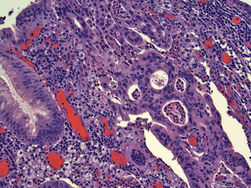
case 1 shows the early lesional area Image
Heavily inflamed mucosa showing solid tumor nodules (left) as well as as trabecular cords of malignant cells with lumen formation.
deeper area of tumor blowing through wall Image
another area Image
case 2 - has diffrent areas including these compact nodules of tuomr, almost looks neuroendocrine (but synapto and chromo are neg) Image
here is has more typical cording of epithelioid cells Image
small focal area shows intestinal appearing type glands Image
tumor si negative for cdx2 - mod path paper shows progressive loss of many gastric cas as they progress through dysplasia into carcinoma for this marker Image
atrophic glands stain for keratin 7, otherwise tumor is actually negative
stains for keratin 20 including normal glands -- a different mod path paper finds about 40% of gastric ca are keratin 7 negative and keratin 20 positive Image
this same tumor appears more like the first one, more classic for intestinal variant, in its metastatic focus in the bone. Image
another tumor shows surface lesion Image
and here it is dissecting through the gastric muscularis Image
polypoid mass Image
hemorrhage Image
The prognosis depends on the stage of the disease at the time of diagnosis and treatment. Early gastric cancer, limited to the mucosa and submucosa, is best treated surgically and has a five-year survival rate of 70-95%. There is no standard chemotherapy for metastatic disease, although the regimen of epirubicin, cisplatin and fluorouracil is the most used regimen, with a median survival of 7-9 months.
• Stomach : Signet Ring Adenocarcinoma
• Stomach : Signet Ring Adenocarcinoma
)
)
)
)
)
)
)
)
)
)
)
)
)
)
)