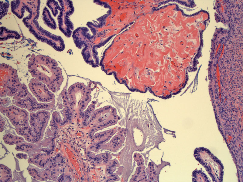

The top image resembles a serous borderline tumor and the bottom image contains papillae lined by indifferent cells (pink polygonal cells with eosinophilic cytoplasm).
Some fibrovascular cores are covered by an admixture of serous and mucinous appearing epithelium. Note the intracytoplasmic mucin vacuoles in some of the mucinous cells. This characteristic blend of serous and mucinous features is the reason some authors champion the term seromucinous tumors of the ovary.
Large dilated glands lined by typical endocervical mucinous cells are seen in the ovarian stroma. The ovarian surface (on the right) is involved by the borderline seromucinous tumor.
The ovarian stroma contains scattered bland glands and psammoma (calcified) bodies. This is not stromal invasion as there is no desmoplastic response.
Areas of the tumor resemble a serous adenofibroma with bland serous cells lining bulbous stroma structures.
At low power, part of a mucinous gland can be seen at the top, with surface involvement of ovary consisting of complex papillary structures
Areas of this tumor are virtually identical to that seen in serous borderline tumors with papillary structures and epithelial tufting.
A closer view demonstrates piled up epithelium with cellular budding. Mucinous cells with bluish mucin can be appreciated in the epithelial lining.
Also known as seromucinous borderline tumor of the ovary or endocervical-like borderline mucinous tumor of mixed cell types, these mucinous ovarian epithelial tumors are composed of endocervical-like mucinous epithelium as well as a mixture of serous, endometrioid, squamous, hobnail and pink polygonal 'indifferent' cells. A more detailed explanation is available in our other case of this entity.
• Ovary : Mucinous Borderline Tumor, Intestinal-type
• Ovary : Mucinous Cystadenoma
• Ovary : Mucinous Borderline Tumor, Endocervical Type
• Ovary : Mucinous Cystadenocarcinoma (Expansile Type)