System: Gynecological: Ovary: Neoplastic: Serous Adenocarcinoma, High Grade Papillary

System: Gynecological: Ovary: Neoplastic: Serous Adenocarcinoma, High Grade Papillary

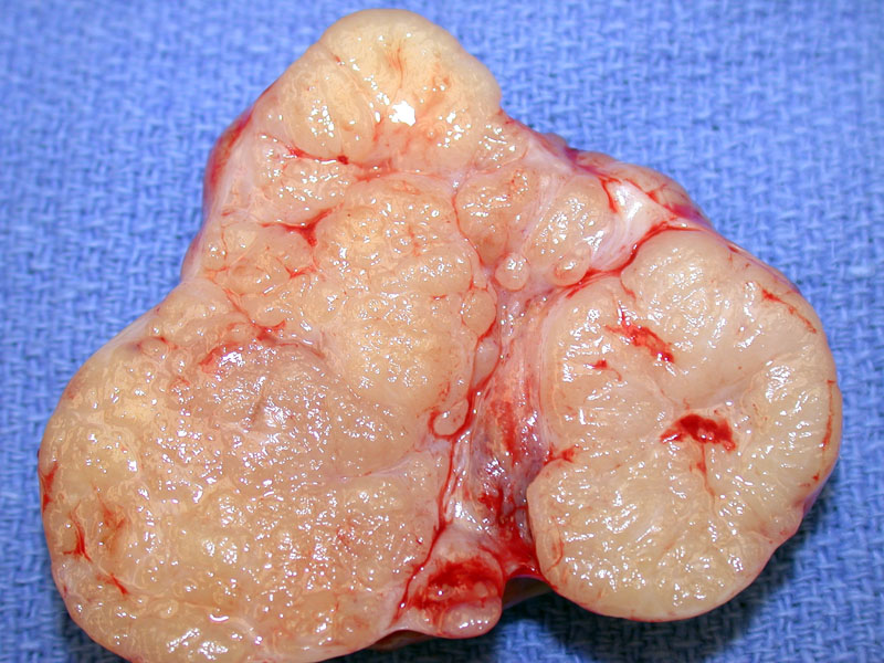
This serous ovarian carcinoma was bisected to reveal a micronodular/papillary cut surface. Fibrous septations are evident.
Papillary structures lined by pseudostratified epithelial cells can be appreciated. Such areas that resemble serous borderline tumor are present at least focally in a high grade serous carcinoma.
In this image, papillary fronds are cut en face to demonstrate scattered bizarre nuclei admixed with other nuclei with high grade atypia.
Neoplastic glands and papillae forming slit-like spaces are highly characteristic.
Another area with papillae and detached cellular buds.
Solid areas of tumor with high grade cellular features invade into the ovarian stroma.
Cystic spaces can form.
Metastatic implants are quite commmon in serous carcinomas. This image demonstrates an implant on pericolonic fat
Metastatic implants can be located on any peritoneal surface -- here is an implant on the uterine serosa, which has invaded into myometrium (bottom image)
Here is an example of a case where the colon had extensive involvement by metastatic high grade serous carcinoma. Note the presence of normal colonic glands (upper right).
MOC 31 of this case, since ddx included colon ca Image
The tumor cells were negative for CEA (CEA is positive in colon adenocarcinoma).
The tumor cells were positive for ER, which further corroborates an ovarian primary.
Other tumors show loss of ER, as in this example in which only this field shows any substantial staining in a minority of the nuclei. Image
Ovarian serous carcinomas are usually CK7 positive and CK20 negative.
WT1 is also a helpful staining in distinguishing between a primary ovarian serous carcinoma (WT1 positive) versus an endometrioid ovarian carcinoma (WT1 negative) or metastatic colorectal carcinoma (WT1 negative). In the colon, the invasive tumor was WT1 positive.
WT1 was also positive on primary ovarian tumor
The omentum demonstrated carcinomatosis -- not an uncommon finding with this malignancy.
Tumor was also identified in the lymph nodes.
Serous tumors as a group account for approximately 30% of all ovarian tumors. Serous carcinomas comprise nearly 50% of malignant tumors of the ovary.
Grossly, the tumor is solid and cystic with foci of necrosis and hemorrhage. The carcinoma frequently invades through the capsule and thus, papillary excrescences are found on the ovarian surface. Microscopically, papillary structures with similar morphology to a borderline tumor are at least focally seen. Solid growth with glands and papillae form characteristic slit-like spaces. Invasion of the stroma is seen as jagged glands, papillae or nests infiltrating a reactive, desmoplastic stroma. Psammoma calcifications are also frequently found.
Most serous carcinomas are high-grade tumors, with obvious nuclear atypia, brisk mitotic activity and solid growth. A minority of serous carcinomas are low-grade and they appear to follow a different pathway than high-grade serous carcinomas. Low-grade carcinomas arise from borderline serous tumors, like borderline tumors, exhibit BRAF, K-RAS or ERBB2 mutations. On the other hand, high-grade serous carcinomas exhibit p53 mutations and do not exhibit BRAF, K-RAS or ERBB2 mutations A precursor lesion to high-grade ovarian carcinomas have not been clearly identified, although microscopic foci of high-grade serous carcinomas have been found, leading many to theorize that high-grade serous carcinomas arise de novo.
Incidence peaks in the 6th and 7th decades (mean age of 56 years). Unfortunately, many patients present with advanced-stage disease. Symptoms may be nonspecific and vague including early satiety, a pelvic or abdominal mass and bloating. Uncommonly, patients will present with an acute onset of abdominal pain due to torsion. Elevated CA-125 is found in ~85% of patients.2
Patients with BRCA1 or BRCA2 mutations with ovarian tumors usually have serous carcinomas, with other common types (clear cell, mucinous) are quite rare in this subgroup of women.1,2
Treatment involves comprehensive surgical staging and an attempt at optimal cytoreduction. Platinum/taxane-based adjuvant chemotherapy should be considered for those with both early- and advanced-stage patients. Careful long-term surveillance is indicated due to the potential for recurrence.
• Ovary : Serous Borderline Tumor
• Ovary : Serous Borderline Tumor, Micropapillary Type
• Fallopian Tube : Papillary Serous Adenocarcinoma
1 Nucci 407-412.
2 Fletcher 573-4.
Boruta DM 2nd, Gehrig PA, Fader AN, Olawaiye AB. Management of women with uterine papillary serous cancer: a Society of Gynecologic Oncology (SGO) review. Gynecol Oncol. 2009 Oct;115(1):142-53. Epub 2009 Jul 9.
Ovarian Low-grade and High-grade Serous Carcinoma: Pathogenesis, Clinicopathologic and Molecular Biologic Features, and Diagnostic Problems
Vang, Russell MD; Shih, Ie-Ming MD, PhD; Kurman, Robert J. MD
Advances in Anatomic Pathology:
September 2009 - Volume 16 - Issue 5 - pp 267-282
)
)
)
)
)
)
)
)
)
)
)
)
)
)
)
)
 were also WT1 positive.', 'special', ''))
)
)