
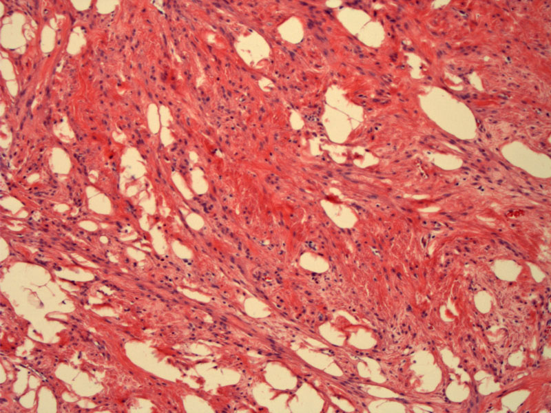
An admixture of smooth muscle cells and adipocytes can be appreciated.
)
Note the lack of cytologic atypia and lack of mitotic activity.
)
This rare variant of uterine leiomyoma is characterized by the presence of adipocytes admixed with smooth muscle cells. Depending on the amount of adipocytes, the cut surface may be quite yellow. Lipoleiomyomas occur in an older age group (post-menopausal) when compared to conventional leiomyomas (reproductive age).1
In a recent study of 50 lipoleiomyomas, the average age was 54. The average tumor size was 4.6 cm. The study found that these tumors do not demonstrate mitotic activity, cytologic atypia, necrosis or calcification. Due to the lack of degenerate changes, the authors concluded that lipoleiomyomas are leiomyomas with adipocyte differentiation rather than fatty degeneration. Follow-up of these cases also confirmed their benign nature.2
A possible differential diagnosis would be a well-differentiated liposarcoma, however, lipoleiomyomas do not exhibit lipoblasts, cytologic atypia or necrosis.2
Benign neoplasm.
1 Fletcher CDM, ed. Diagnostic Histopathology of Tumors. 3rd Ed. Philadelphia, PA: Elsevier; 2007: 686-7.
2 Wang X, Kumar D, Seidman JD. Uterine lipoleiomyomas: a clinicopathologic study of 50 cases. Int J Gynecol Pathol. 2006 Jul;25(3):239-42.