
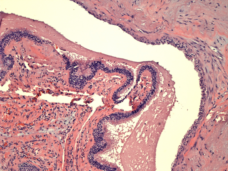
The cyst shows a simple epithelial lining and is filled with luminal hypocellular serous fluid.
)
ciliated columnar epithelium lines this simple esophageal cyst. The cyst lining can vary and include the possibility of squamous, columnar, cuboidal, pseudostratified, ciliated, and gastric mucosal lining.
)
FNA consists largely of histiocytes obtained from the cyst contents.
)
Simple cysts involve duplication of the epithelium, whereas true esophageal duplications are comprised of the submucosa and the muscle wall without duplication of the epithelium. Maldevelopment of the posterior division of the primitive foregut causes esophageal cysts. These cysts do not communicate with the lumen of the esophagus.
Most esophageal cysts are asymptomatic when detected. Chest pain or dysphagia may occasionally be presenting symptom(s).
Most should be resected because up to 75% may evenutually become symptomatic.