
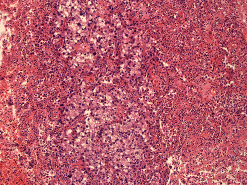
A cluster of clear cells surrounded by inflammatory debris in this acutely inflamed biopsy material. Grossly, the lesion can be polypoid and likely in this case, ulcerated. Image
)
A closer look demonstrates a solid growth of cells with abundant clear cytoplasm. The nuclei are clearly atypical and hyperchromatic.
)
Brisk mitotic activity is usually seen. There is a mitotic figure at about 9 o clock. Can you find it?
)
The clear cells are forming loose nests with cystic spaces filled by eosinophilic secretions.
)
Clear cell adenocarcinoma of the vagina is most famously associated with DES exposure in utero. However, this malignancy can also occur sporadically. DES exposure also leads to other abnormalities in the female genital tract such as vaginal adenosis and cervical ectropion. Vaginal adenosis is often seen adjacent to invasive clear cell carcinoma and may be a precursor lesion (Fletcher).
Microscopically, clear cells with abundant glycogen are arranged in tubulocystic, solid or papillary patterns. The nuclei are pleomorphic. Mitotic activity is brisk. Areas of adenosis may be found adjacent to the tumor.
Estimated risk if DES exposed = 1 per 1000 and age of detection peaks at age 20. Extremely rare in those without in utero DES exposure and usually arises as a post-menopausal malignancy. Typically presents with abnormal vaginal bleeding or dyspareunia. There may be a clinically evident a polypoid or nodular mass.
Treatment involves radical hysterectomy with lymphadenectomy and concurrent chemoradiation if the lesion is restricted to the upper third of the vagina. Total vaginectomy or even exenteration may be necessary depending on extent of the lesion.
Stage is the most important prognostic factor. Overall, outcome is very good with 5 and 10 year survival rates of 91 and 85% respectively (Nucci).
• Cervix : Clear Cell (Adeno)carcinoma
Fletcher CDM, ed. Diagnostic Histopathology of Tumors. 3rd Ed. Philadelphia, PA: Elsevier; 2007: 720-1.
Nucci MR, Oliva Esther. Gynecologic Pathology: Foundations in Diagnostic Pathology. Philadelphia, PA: Elsevier: 2009: 119-121.
***Images courtesy of Dr. Lida Crooks, VAMC, Albuquerque NM. Case materials from 1978.