System: Head and Neck: Oral Cavity: Neoplastic: OVERRIDE

System: Head and Neck: Oral Cavity: Neoplastic: OVERRIDE

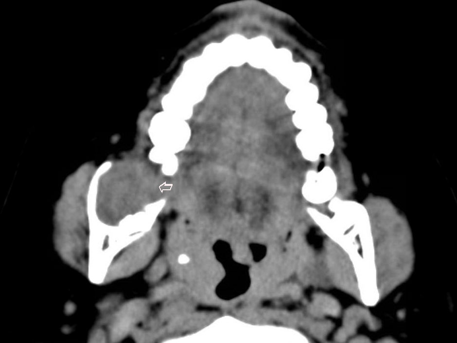
CT scan shows a 3 cm right mandibular mass.
Case 3: The classic follicular pattern is well-demonstrated here with follicles composed of ameloblastic epithelium. Inside the follicles, a loose network of stellate cells are seen.
A more plexiform pattern is seen in this area. The neoplastic cells form interconnecting cords and the stroma is quite collagenous and dense, although not quite desmoplastic.
More than one pattern is often encountered in an ameloblastoma as seen here. The top part of the image is plexiform/desmoplastic whereas the lower half of the image demonstrates an ill-defined follicles surrounded by a spindled stroma.
)
)
)
)