
Case 1: There is usually a central elongated duct surrounded by tubules. The tubules conmmonly contain eosinophilic secretions resembling thyroid colloid. Mesonephric remnants are usually located deep within the cervical stroma, however, they can occasionally be found toward the surface and even blend in with endocervical glandular clefts (Mills).
)
The tubules (seen clearly on the lower right) are lined by bland cuboidal cells with centrally placed round nuclei. Even though these tubular structures may be deep within the stroma, they do not have the ragged infiltrative structures of endocervical carcinoma (an important distinguishing feature).
)
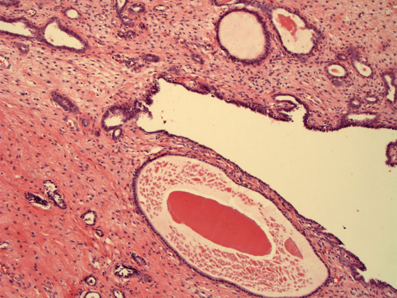
Case 2: A central duct is surrounded by tubules.
)
A closer look at the duct and tubules.
)
The tubules are lined by cuboidal, bland-appearing cells.
)
Case 3: Small tubules percolate through the cervical stroma. You can see how it would be important to recognize this entity.
)
Eosinophilic secretions are often seen in the lumina. Again, the lining cells are bland.
)
Yet another image of the rounded mesonephric tubules.
)
Mesonephric remnants are formed from the Wolffian duct as it regresses during the development of the female reproductive tract. These vestigial structures persist as Gartner duct cysts in the vagina and mesonephric remnants in the cervix. Mesonephric remnants can be found in up to 1/3 of women (Mills).
In the cervix, mesonephric remannts are important to recognize as they can mimic well-differentiated adenocarcinoma of the cervix, especially if the remnants become hyperplastic, termed mesonephric hyperplasia. A benign cytologic appearance, intraluminal eosinophilic secretions, lack of desmoplastic stromal response, lack of involvement of the overlying squamous epithelium, orderly architectural pattern are helpful features to distinguish mesonephric rests or mesonephric hyperplasia from well-differentiated endocervical adenocarcinoma. Rarely, these structures evolve into mesonephric adenocarcinoma. Note that mesonephric rests and mesonephric adenocarcinoma stain positively with CD10, a useful diagnostic tool (Sternberg).
• Ovary : Mesonephric Adenocarcinoma from Cervix
• Cervix : Mesonephric Adenocarcinoma
Mills, S. Histology for Pathologists. 3rd Ed. Philadelphia, PA: Lippincott, Williams & Wilkins; 2007: 1025-6.
Sternberg SS, ed. Diagnostic Surgical Pathology.4th Ed. Philadelphia, PA: Lippincott Williams & Wilkins; 2004: 2405-6.