System: Gastrointestinal: Gallbladder: Benign: Adenoma

System: Gastrointestinal: Gallbladder: Benign: Adenoma

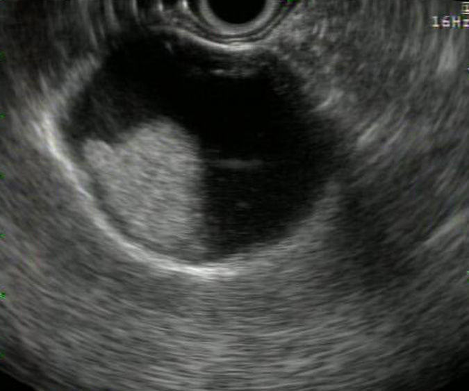
Ultrasound of gallbladder illustrates a large polypoid intraluminal pedunculated mass.
Case 1: Variably-sized glands are separated by a loose stroma.
The glands are lined by adenomatous epithelium. Dysplasia can be seen in gallbladder adenomas, although the adenoma-dysplasia-carcinoma has not yet been fully defined in this organ.
Fronds lined by normal epithelium showing cholesterolosis (left) abuts the adenoma (right).
In this area, there are dilated glands filled with eosinophilic material.
Case 2: Another gallbladder adenoma shows variably sized tubular glands and areas of hemorrhage.
The epithelium lining the tubules can be pyloric-type (resembling pyloric glands), intestinal-type (resembling colonic adenomas) or biliary type (rare, resembling normal gallbladder). In this case, the lining most resembles pyloric type glands, although they are some what cuboidal.
Adenomas of the gallbladder are rare lesions and for the most part benign. These findings are usually less than 2 cm in size and solitary. Microscopically, tubular glands are separated by stroma, which may be fibrous, hyalinized or edematous. The epithelium lining the glands may be pyloric (most common) or intestinal type (Fletcher).
Although the majority of these neoplasms are benign, dysplastic changes and even carcinoma-in-situ can be seen in some instances. As such, these are now considered a premalignant lesion with the risk of malignancy increasing with size and degree of papilla formation. An adenoma-dysplasia-carcinoma sequence similar to that in the colon has not been fully elucidated in the gallbladder, however.
Predominately affects females. Presents with symptoms similar to cholecystitis. Rarely, patients also have associated Peutz-Jegher or Gardner syndrome.
Usually benign and cured by excision.
Cheng L, Bostwick DG, eds. Essentials of Anatomic Pathology. 2nd Ed. Totowa, NJ: Humana Press; 2006: 1434.
Fletcher CDM, ed. Diagnostic Histopathology of Tumors. 3rd Ed. Philadelphia, PA: Elsevier; 2007: 444.
**Case 2 contributed by Dr. Kate Sciandra, Dept of Pathology, VAMC Albuquerque New Mexico.
)
)
)
)
)
)
)